File:PMC3883063 hr-2013-4-e16-g002.png
Jump to navigation
Jump to search
Size of this preview: 415 × 599 pixels. Other resolutions: 166 × 240 pixels | 488 × 704 pixels.
Original file (488 × 704 pixels, file size: 321 KB, MIME type: image/png)
Summary
| Description |
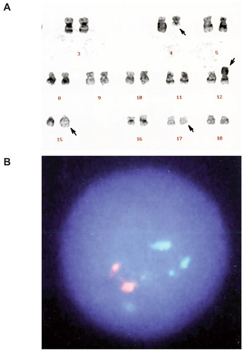
English: fig002: A) G-banded karyotype of the bone marrow cells showing t(4;12)(q21;p11) and t(15;17)(q22;q21). Arrows indicate the derivative chromosomes. B) FISH analysis with PML/RARα-specific probes showing two orange (PML) and two green (RARα) signals. No PML/RARα fusion signal (which should appear yellow) was detected. |
| Date | |
| Source | https://openi.nlm.nih.gov/detailedresult?img=PMC3883063_hr-2013-4-e16-g002&query=&req=4 |
| Author | Saito M, Izumiyama K, Mori A, Irie T, Tanaka M, Morioka M, Musashi M |
Licensing
English: This file is licensed CC BY-NC 3.0
This file was uploaded with UploadWizard.
File history
Click on a date/time to view the file as it appeared at that time.
| Date/Time | Thumbnail | Dimensions | User | Comment | |
|---|---|---|---|---|---|
| current | 20:46, 23 November 2022 | | 488 × 704 (321 KB) | Ozzie10aaaa (talk | contribs) | Uploaded a work by Saito M, Izumiyama K, Mori A, Irie T, Tanaka M, Morioka M, Musashi M from https://openi.nlm.nih.gov/detailedresult?img=PMC3883063_hr-2013-4-e16-g002&query=&req=4 with UploadWizard |
You cannot overwrite this file.
File usage
There are no pages that use this file.