File:PMC3921946 CRIM.PEDIATRICS2014-256356.001.png
PMC3921946_CRIM.PEDIATRICS2014-256356.001.png (512 × 216 pixels, file size: 169 KB, MIME type: image/png)
License
Attribution 3.0 Unported (CC BY 3.0)
Summary
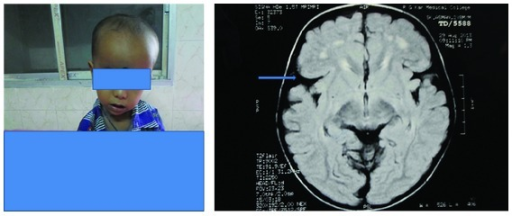
Author:Pusti S, Das N, Nayek K, Biswas S, Department of Paediatrics, R. G. Kar Medical College and Hospital, Khudiram Bose Sarani, Kolkata (Openi/National Library of Medicine) Source:https://openi.nlm.nih.gov/detailedresult?img=PMC3921946_CRIM.PEDIATRICS2014-256356.001&query=Glutaric%20aciduria%20type%201&it=xg&req=4&npos=3 Description:fig1: Showing patient with macrocephaly, typical facies, and MRI of his brain reveals frontotemporal atrophy, dilated sylvian fissures with open opercula (arrow), diffuse hyperintense lesions in bilateral basal ganglia, and both frontal white matter and bilateral periventricular area. Widening of the sylvian fissure gives the characteristic “bat-wing” appearance.
File history
Click on a date/time to view the file as it appeared at that time.
| Date/Time | Thumbnail | Dimensions | User | Comment | |
|---|---|---|---|---|---|
| current | 21:25, 16 August 2021 | | 512 × 216 (169 KB) | Ozzie10aaaa (talk | contribs) | Author:Pusti S, Das N, Nayek K, Biswas S, Department of Paediatrics, R. G. Kar Medical College and Hospital, Khudiram Bose Sarani, Kolkata (Openi/National Library of Medicine) Source:https://openi.nlm.nih.gov/detailedresult?img=PMC3921946_CRIM.PEDIATRICS2014-256356.001&query=Glutaric%20aciduria%20type%201&it=xg&req=4&npos=3 Description:fig1: Showing patient with macrocephaly, typical facies, and MRI of his brain reveals frontotemporal atrophy, dilated sylvian fissures with open opercula (arro... |
You cannot overwrite this file.
File usage
There are no pages that use this file.