File:PMC3949000 kjp-52-85-g001.png
PMC3949000_kjp-52-85-g001.png (512 × 121 pixels, file size: 170 KB, MIME type: image/png)
License
Attribution-NonCommercial 3.0 Unported (CC BY-NC 3.0)
Summary
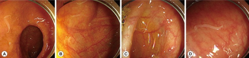
Author:Kim BJ, Song KS, Kong HH, Cha HJ, Ock M,Department of Internal Medicine, On Hospital(Openi/National Library of Medicine) Source:https://openi.nlm.nih.gov/detailedresult?img=PMC3949000_kjp-52-85-g001&query=Hymenolepiasis&it=xg&req=4&npos=9 Description:F1: Patient colonoscopy findings. Colonoscopy revealed that a large number of Hymenolepis nana adult worms were scattered throughout the colon as well as in the terminal ileum. (A) Terminal ileum. (B) Cecum. (C) Transverse colon. (D) Sigmoid colon.
File history
Click on a date/time to view the file as it appeared at that time.
| Date/Time | Thumbnail | Dimensions | User | Comment | |
|---|---|---|---|---|---|
| current | 22:05, 16 December 2021 | 512 × 121 (170 KB) | Ozzie10aaaa (talk | contribs) | Author:Kim BJ, Song KS, Kong HH, Cha HJ, Ock M,Department of Internal Medicine, On Hospital(Openi/National Library of Medicine) Source:https://openi.nlm.nih.gov/detailedresult?img=PMC3949000_kjp-52-85-g001&query=Hymenolepiasis&it=xg&req=4&npos=9 Description:F1: Patient colonoscopy findings. Colonoscopy revealed that a large number of Hymenolepis nana adult worms were scattered throughout the colon as well as in the terminal ileum. (A) Terminal ileum. (B) Cecum. (C) Transverse colon. (D) Sigmo... |
You cannot overwrite this file.
File usage
There are no pages that use this file.