File:PMC3973996 1129-2377-15-13-3.png
PMC3973996_1129-2377-15-13-3.png (512 × 491 pixels, file size: 236 KB, MIME type: image/png)
License
[Attribution 2.0 Generic (CC BY 2.0)]
Summary
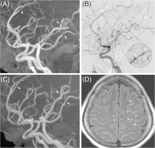
Author:Cheng YC, Kuo KH, Lai TH,Section of Neurology, Department of Internal Medicine, Far Eastern Memorial Hospital(Openi/National Library of Medicine)Source:https://openi.nlm.nih.gov/detailedresult?img=PMC3973996_1129-2377-15-13-3&query=Reversible%20cerebral%20vasoconstriction%20syndrome&it=xg&req=4&npos=1 Description:F3: Imaging findings of reversible cerebral vasoconstriction syndrome. Multifocal vasoconstriction demonstrated by magnetic resonance angiography (MRA) (A) and catheter angiography (B), involving the anterior, middle, and posterior cerebral arteries. Follow-up MRA (C) revealed significant interval resolution of the previous lesions. Axial fluid attenuated inversion recovery (FLAIR) imaging revealed linear hyperintensity lesions in the sulci of the bilateral frontal lobes (D).
File history
Click on a date/time to view the file as it appeared at that time.
| Date/Time | Thumbnail | Dimensions | User | Comment | |
|---|---|---|---|---|---|
| current | 20:51, 31 January 2022 | | 512 × 491 (236 KB) | Ozzie10aaaa (talk | contribs) | Author:Cheng YC, Kuo KH, Lai TH,Section of Neurology, Department of Internal Medicine, Far Eastern Memorial Hospital(Openi/National Library of Medicine)Source:https://openi.nlm.nih.gov/detailedresult?img=PMC3973996_1129-2377-15-13-3&query=Reversible%20cerebral%20vasoconstriction%20syndrome&it=xg&req=4&npos=1 Description:F3: Imaging findings of reversible cerebral vasoconstriction syndrome. Multifocal vasoconstriction demonstrated by magnetic resonance angiography (MRA) (A) and catheter angiog... |
You cannot overwrite this file.
File usage
There are no pages that use this file.