File:PMC3975201 cde-0006-0037-g03.png
PMC3975201_cde-0006-0037-g03.png (512 × 309 pixels, file size: 340 KB, MIME type: image/png)
License
Attribution-NonCommercial 3.0 Unported (CC BY-NC 3.0)
Summary
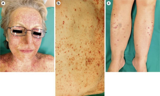
Author:Kuonen F, Bucher M, de Leval L, Vernez M, Gilliet M, Conrad C, Feldmeyer L , Department of Dermatology and Venereology, Hôpital de Beaumont, Lausanne University Hospital Center(Openi/National Library of Medicine) Source:https://openi.nlm.nih.gov/detailedresult?img=PMC3975201_cde-0006-0037-g03&query=Hepatosplenic%20T-cell%20lymphoma&it=xg&req=4&npos=23 Description:F3: Photographs of the clinical evolution 2 months after the initial presentation. The clinical photographs show an intensification of the purpuric eruption of the face (a) as well as an extension of the purpura to the upper chest (b) and legs (c).
File history
Click on a date/time to view the file as it appeared at that time.
| Date/Time | Thumbnail | Dimensions | User | Comment | |
|---|---|---|---|---|---|
| current | 19:47, 11 August 2021 | | 512 × 309 (340 KB) | Ozzie10aaaa (talk | contribs) | Author:Kuonen F, Bucher M, de Leval L, Vernez M, Gilliet M, Conrad C, Feldmeyer L , Department of Dermatology and Venereology, Hôpital de Beaumont, Lausanne University Hospital Center(Openi/National Library of Medicine) Source:https://openi.nlm.nih.gov/detailedresult?img=PMC3975201_cde-0006-0037-g03&query=Hepatosplenic%20T-cell%20lymphoma&it=xg&req=4&npos=23 Description:F3: Photographs of the clinical evolution 2 months after the initial presentation. The clinical photographs show an intensif... |
You cannot overwrite this file.
File usage
There are no pages that use this file.