File:PMC3990818 kjpain-27-112-g001 (1).png
PMC3990818_kjpain-27-112-g001_(1).png (512 × 190 pixels, file size: 103 KB, MIME type: image/png)
License
Attribution-NonCommercial 3.0 Unported (CC BY-NC 3.0)
Summary
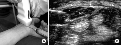
Author:Kim HJ, Park SH, Department of Anesthesiology and Pain Medicine, Jeju National University School of Medicine(Openi/National Library of Medicine) Source:https://openi.nlm.nih.gov/detailedresult?img=PMC3990818_kjpain-27-112-g001&query=Carpal%20tunnel%20syndrome&it=xg&req=4&npos=5 Description:F1: (A) Ultrasound-guided carpal tunnel injection. It shows transducer position for transverse imaging of the carpal tunnel and in-plane needle approach. (B) Transverse sonogram of the left carpal tunnel in a patient with carpal tunnel syndrome. Arrows indicate flexor retinaculum, Asterisk: anechoic injectate, N: needle, MN: median nerve, T: flexor tendons (These figures are quoted from the paper of Smith et al. [19] after permission.).
File history
Click on a date/time to view the file as it appeared at that time.
| Date/Time | Thumbnail | Dimensions | User | Comment | |
|---|---|---|---|---|---|
| current | 22:22, 13 December 2021 | 512 × 190 (103 KB) | Ozzie10aaaa (talk | contribs) | Author:Kim HJ, Park SH, Department of Anesthesiology and Pain Medicine, Jeju National University School of Medicine(Openi/National Library of Medicine) Source:https://openi.nlm.nih.gov/detailedresult?img=PMC3990818_kjpain-27-112-g001&query=Carpal%20tunnel%20syndrome&it=xg&req=4&npos=5 Description:F1: (A) Ultrasound-guided carpal tunnel injection. It shows transducer position for transverse imaging of the carpal tunnel and in-plane needle approach. (B) Transverse sonogram of the left carpal tu... |
You cannot overwrite this file.
File usage
There are no pages that use this file.
.png&oldid=1237620)