File:PMC4017419 GHFBB-4-164-g001.png

Original file (512 × 716 pixels, file size: 940 KB, MIME type: image/png)
License
Attribution-NonCommercial 3.0 Unported (CC BY-NC 3.0)
Summary
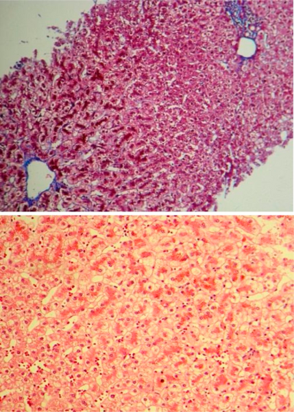
Author:Lahmi F, Roshani M, Khosravi K, Azizi M, Mohebbi SR, Zali MR ,Research Institute for Gastroenterology and Liver Disease, Shahid Beheshti University of Medical sciences (Openi/National Library of Medicine) Source:https://openi.nlm.nih.gov/detailedresult?img=PMC4017419_GHFBB-4-164-g001&query=Dubin%E2%80%93Johnson%20syndrome&it=xg&req=4&npos=2 Description:F0001: Histopathological view of liver needle biopsy shows intact lobular and vascular architecture. Individual hepatocytes contain abundant coarse brown pigment granules especially in perivenular areas. Portal tracts show mild lymphocytic infiltration.
File history
Click on a date/time to view the file as it appeared at that time.
| Date/Time | Thumbnail | Dimensions | User | Comment | |
|---|---|---|---|---|---|
| current | 15:43, 12 October 2021 | | 512 × 716 (940 KB) | Ozzie10aaaa (talk | contribs) | Author:Lahmi F, Roshani M, Khosravi K, Azizi M, Mohebbi SR, Zali MR ,Research Institute for Gastroenterology and Liver Disease, Shahid Beheshti University of Medical sciences (Openi/National Library of Medicine) Source:https://openi.nlm.nih.gov/detailedresult?img=PMC4017419_GHFBB-4-164-g001&query=Dubin%E2%80%93Johnson%20syndrome&it=xg&req=4&npos=2 Description:F0001: Histopathological view of liver needle biopsy shows intact lobular and vascular architecture. Individual hepatocytes contain abu... |
You cannot overwrite this file.
File usage
There are no pages that use this file.