File:PMC4056107 fimmu-05-00281-g001.png
PMC4056107_fimmu-05-00281-g001.png (512 × 130 pixels, file size: 166 KB, MIME type: image/png)
License
Attribution 3.0 Unported (CC BY 3.0)
Summary
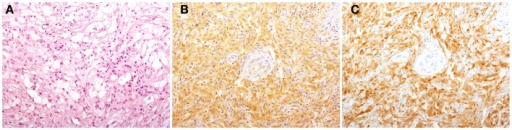
Author:Cavalli G, Biavasco R, Borgiani B, Dagna L , Unit of Internal Medicine and Clinical Immunology, IRCCS San Raffaele Scientific Institute (Openi/National Library of Medicine) Source:https://openi.nlm.nih.gov/detailedresult?img=PMC4056107_fimmu-05-00281-g001&query=Erdheim%E2%80%93Chester%20disease&it=xg&req=4&npos=2 Description:F1: Histological findings in patients with Erdheim–Chester disease (ECD). Histology shows a xanthogranulomatous infiltrate composed by foamy histiocytes accompanied by fibrosis [(A), H&E, original magnification 200×]. Immunohistochemical studies reveal that some of the infiltrating histiocytes stain for BRAFV600E [(B), VE1 immunostaining, 200×], and p16Ink4a [(C), p16Ink4a immunostaining, 200×].
File history
Click on a date/time to view the file as it appeared at that time.
| Date/Time | Thumbnail | Dimensions | User | Comment | |
|---|---|---|---|---|---|
| current | 20:59, 24 October 2021 | 512 × 130 (166 KB) | Ozzie10aaaa (talk | contribs) | Author:Cavalli G, Biavasco R, Borgiani B, Dagna L , Unit of Internal Medicine and Clinical Immunology, IRCCS San Raffaele Scientific Institute (Openi/National Library of Medicine) Source:https://openi.nlm.nih.gov/detailedresult?img=PMC4056107_fimmu-05-00281-g001&query=Erdheim%E2%80%93Chester%20disease&it=xg&req=4&npos=2 Description:F1: Histological findings in patients with Erdheim–Chester disease (ECD). Histology shows a xanthogranulomatous infiltrate composed by foamy histiocytes accompanie... |
You cannot overwrite this file.
File usage
There are no pages that use this file.