File:PMC4105805 kjr-15-439-g001.png
PMC4105805_kjr-15-439-g001.png (512 × 263 pixels, file size: 217 KB, MIME type: image/png)
License
Attribution-NonCommercial 3.0 Unported (CC BY-NC 3.0)
Summary
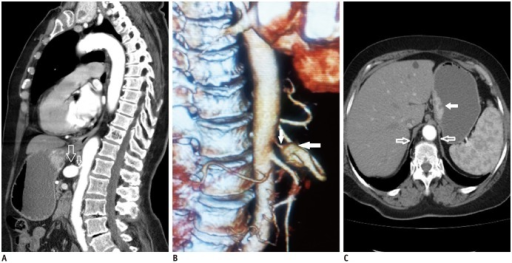
Author:Gunduz Y, Asil K, Aksoy YE, Tatlı Ayhan L,Department of Radiology, Sakarya University Medical Faculty(Openi/National Library of Medicine)Source:https://openi.nlm.nih.gov/detailedresult?img=PMC4105805_kjr-15-439-g001&query=Median%20arcuate%20ligament%20syndrome&it=xg&req=4&npos=1 Description:F1: Median arcuate ligament syndrome in 72-year-old male patient.A. Sagittal reformatted contrast enhanced CT angiography shows stenosis and aneurysm of celiac artery due to compression by median arcuate ligament (arrows). B. Three-dimensional reconstruction CT angiography shows severe stenosis and poststenotic dilatation (white arrows) of celiac artery. C. Axial CT image shows median arcuate ligaments (hollow arrows) and gastric mucasal thickening and contrast enhancement (white arrow).
File history
Click on a date/time to view the file as it appeared at that time.
| Date/Time | Thumbnail | Dimensions | User | Comment | |
|---|---|---|---|---|---|
| current | 23:30, 16 January 2022 | | 512 × 263 (217 KB) | Ozzie10aaaa (talk | contribs) | Author:Gunduz Y, Asil K, Aksoy YE, Tatlı Ayhan L,Department of Radiology, Sakarya University Medical Faculty(Openi/National Library of Medicine)Source:https://openi.nlm.nih.gov/detailedresult?img=PMC4105805_kjr-15-439-g001&query=Median%20arcuate%20ligament%20syndrome&it=xg&req=4&npos=1 Description:F1: Median arcuate ligament syndrome in 72-year-old male patient.A. Sagittal reformatted contrast enhanced CT angiography shows stenosis and aneurysm of celiac artery due to compression by median ar... |
You cannot overwrite this file.
File usage
There are no pages that use this file.