File:PMC4129610 LI-31-296-g001.png
Jump to navigation
Jump to search
No higher resolution available.
PMC4129610_LI-31-296-g001.png (512 × 209 pixels, file size: 102 KB, MIME type: image/png)
Summary
| Description |
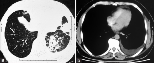
English: (a) Contrast enhanced computed tomography chest axial image in lung window setting show areas of consolidations in inferior segment of lingular lobe and lateral and posterior basal segments of left lower lobe (b) Axial image in mediastinal window demonstrate left pleural effusion |
| Date | |
| Source | https://openi.nlm.nih.gov/detailedresult?img=PMC4129610_LI-31-296-g001&query=Selective%20IgM%20immunodeficiency&it=xg&req=4&npos=3 |
| Author | Dhir V, Sagar V, Aggarwal A, Rawat A, Singhal M |
Licensing
English: This file is licensed CC BY-NC-SA 3.0
This file was uploaded with UploadWizard.
File history
Click on a date/time to view the file as it appeared at that time.
| Date/Time | Thumbnail | Dimensions | User | Comment | |
|---|---|---|---|---|---|
| current | 18:37, 11 May 2022 | 512 × 209 (102 KB) | Ozzie10aaaa (talk | contribs) | Uploaded a work by Dhir V, Sagar V, Aggarwal A, Rawat A, Singhal M from https://openi.nlm.nih.gov/detailedresult?img=PMC4129610_LI-31-296-g001&query=Selective%20IgM%20immunodeficiency&it=xg&req=4&npos=3 with UploadWizard |
You cannot overwrite this file.
File usage
There are no pages that use this file.