File:PMC4156028 f1000research-3-3493-g0003.png
PMC4156028_f1000research-3-3493-g0003.png (512 × 438 pixels, file size: 483 KB, MIME type: image/png)
License
https://creativecommons.org/licenses/by/4.0/ Attribution 4.0 International (CC BY 4.0)
- &
[CC0 1.0 Universal (CC0 1.0) Public Domain Dedication]
Summary
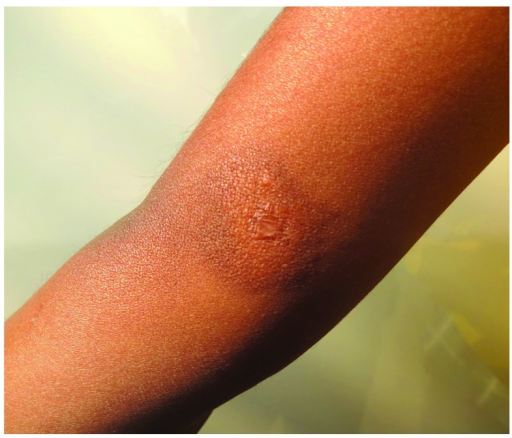
Author:Sukesh MS, Dandale A, Dhurat R, Sarkate A, Ghate S,Dermatology Department, LTM Medical College and General Hospital (Openi/National Library of Medicine) Source:https://openi.nlm.nih.gov/detailedresult?img=PMC4156028_f1000research-3-3493-g0003&query=codeine&it=xg&req=4&npos=55 Description:f4: Complete subsidence of the lesion with residual marginal pigmentation noted at the end of three months of therapy.The central atrophic scar due to biopsy can be seen in the centre of the lesion.
File history
Click on a date/time to view the file as it appeared at that time.
| Date/Time | Thumbnail | Dimensions | User | Comment | |
|---|---|---|---|---|---|
| current | 19:46, 19 September 2021 | | 512 × 438 (483 KB) | Ozzie10aaaa (talk | contribs) | Author:Sukesh MS, Dandale A, Dhurat R, Sarkate A, Ghate S,Dermatology Department, LTM Medical College and General Hospital (Openi/National Library of Medicine) Source:https://openi.nlm.nih.gov/detailedresult?img=PMC4156028_f1000research-3-3493-g0003&query=codeine&it=xg&req=4&npos=55 Description:f4: Complete subsidence of the lesion with residual marginal pigmentation noted at the end of three months of therapy.The central atrophic scar due to biopsy can be seen in the centre of the lesion. |
You cannot overwrite this file.
File usage
There are no pages that use this file.