File:PMC4168642 JCIS-4-48-g002.png
PMC4168642_JCIS-4-48-g002.png (512 × 516 pixels, file size: 173 KB, MIME type: image/png)
License
Attribution-NonCommercial-ShareAlike 3.0 Unported (CC BY-NC-SA 3.0)
Summary
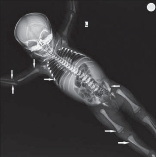
Author:Kalyanasundaram K, Jegadeesan P, Mohan SC, Ponnurangam VN,Department of Pediatrics, Sri Ramachandra Medical College (Openi/National Library of Medicine) Source:https://openi.nlm.nih.gov/detailedresult?img=PMC4168642_JCIS-4-48-g002&query=Malignant%20infantile%20osteopetrosis&it=xg&req=4&npos=1 Description:F1: 45-dayold infant presented with splenohepatomegaly, which was subsequently diagnosed as malignant infantile osteopetrosis. X-ray of the skeleton shows (solid arrow) bone in bone appearance in the femur, tibia, humerus, radius, ulna, and iliac bones.
File history
Click on a date/time to view the file as it appeared at that time.
| Date/Time | Thumbnail | Dimensions | User | Comment | |
|---|---|---|---|---|---|
| current | 22:11, 28 August 2021 | | 512 × 516 (173 KB) | Ozzie10aaaa (talk | contribs) | Author:Kalyanasundaram K, Jegadeesan P, Mohan SC, Ponnurangam VN,Department of Pediatrics, Sri Ramachandra Medical College (Openi/National Library of Medicine) Source:https://openi.nlm.nih.gov/detailedresult?img=PMC4168642_JCIS-4-48-g002&query=Malignant%20infantile%20osteopetrosis&it=xg&req=4&npos=1 Description:F1: 45-dayold infant presented with splenohepatomegaly, which was subsequently diagnosed as malignant infantile osteopetrosis. X-ray of the skeleton shows (solid arrow) bone in bone ap... |
You cannot overwrite this file.
File usage
There are no pages that use this file.