File:PMC4177064 12878 2013 28 Fig2 HTML.png
PMC4177064_12878_2013_28_Fig2_HTML.png (512 × 248 pixels, file size: 261 KB, MIME type: image/png)
License
Attribution 2.0 Generic (CC BY 2.0)
- &
CC0 1.0 Universal (CC0 1.0) Public Domain Dedication
Summary
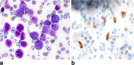
Author:Cehreli C, Alacacioglu I, Piskin O, Ates H, Cehreli R, Calibasi G, Yuksel E, Ozkal S, Ozsan GH,Division of Hematology, Dokuz Eylul University School of Medicine (Openi/National Library of Medicine) Source:https://openi.nlm.nih.gov/detailedresult?img=PMC4177064_12878_2013_28_Fig2_HTML&query=Mast%20cell%20leukemia&it=xg&req=4&npos=4 Description:Fig2: Showing aggregates of mast cells containing mixed black and orange color round cytoplasmic granules and a giant segmented basophil in (a) (wright’s stain × 100), brown color round granular cytoplasmic staining demonstrated by tryptase immunohistochemical staining on PB smear representing mast cells in (b) (×100).
File history
Click on a date/time to view the file as it appeared at that time.
| Date/Time | Thumbnail | Dimensions | User | Comment | |
|---|---|---|---|---|---|
| current | 18:32, 19 February 2022 | | 512 × 248 (261 KB) | Ozzie10aaaa (talk | contribs) | Author:Cehreli C, Alacacioglu I, Piskin O, Ates H, Cehreli R, Calibasi G, Yuksel E, Ozkal S, Ozsan GH,Division of Hematology, Dokuz Eylul University School of Medicine (Openi/National Library of Medicine) Source:https://openi.nlm.nih.gov/detailedresult?img=PMC4177064_12878_2013_28_Fig2_HTML&query=Mast%20cell%20leukemia&it=xg&req=4&npos=4 Description:Fig2: Showing aggregates of mast cells containing mixed black and orange color round cytoplasmic granules and a giant segmented basophil in (a) (... |
You cannot overwrite this file.
File usage
There are no pages that use this file.