File:PMC4192343 12876 2014 1193 Fig1 HTML.png
PMC4192343_12876_2014_1193_Fig1_HTML.png (512 × 256 pixels, file size: 332 KB, MIME type: image/png)
License
Attribution 4.0 International (CC BY 4.0)
- &
CC0 1.0 Universal (CC0 1.0) Public Domain Dedication
Summary
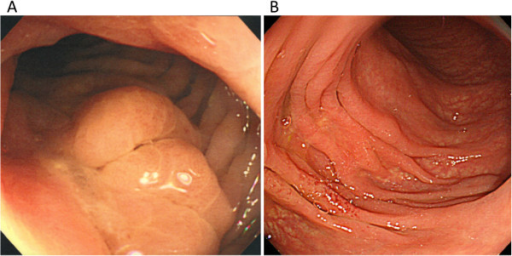
Author:Matsubayashi H, Kishida Y, Yoshida Y, Yoshida M, Tanaka Y, Igarashi K, Imai K, Ono H, Division of Endoscopy, Shizuoka Cancer Center(Openi/National Library of Medicine) Source:https://openi.nlm.nih.gov/detailedresult?img=PMC4192343_12876_2014_1193_Fig1_HTML&query=Retroperitoneal%20fibrosis&it=xg&req=4&npos=99 Description:Fig1: Colonoscopic views at the splenic flexure before (A) and after (B) steroid therapy. The erosion associated with a converging fold and luminal stenosis (A). Healed erosion with an indistinct mucosal vascular pattern (B).
File history
Click on a date/time to view the file as it appeared at that time.
| Date/Time | Thumbnail | Dimensions | User | Comment | |
|---|---|---|---|---|---|
| current | 17:17, 4 January 2022 | | 512 × 256 (332 KB) | Ozzie10aaaa (talk | contribs) | Author:Matsubayashi H, Kishida Y, Yoshida Y, Yoshida M, Tanaka Y, Igarashi K, Imai K, Ono H, Division of Endoscopy, Shizuoka Cancer Center(Openi/National Library of Medicine) Source:https://openi.nlm.nih.gov/detailedresult?img=PMC4192343_12876_2014_1193_Fig1_HTML&query=Retroperitoneal%20fibrosis&it=xg&req=4&npos=99 Description:Fig1: Colonoscopic views at the splenic flexure before (A) and after (B) steroid therapy. The erosion associated with a converging fold and luminal stenosis (A). Healed... |
You cannot overwrite this file.
File usage
There are no pages that use this file.