File:PMC4195840 13244 2014 349 Fig17 HTML.png
Jump to navigation
Jump to search
No higher resolution available.
PMC4195840_13244_2014_349_Fig17_HTML.png (512 × 172 pixels, file size: 144 KB, MIME type: image/png)
Summary
| Description |
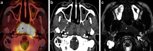
English: Fig17: a Axial PET/CT image reveals a FDG-avid mass in the nasopharynx in keeping with a known nasopharyngeal carcinoma (asterisk). No separate retropharyngeal adenopathy is seen on this image. b Corresponding axial CECT image shows the infiltrative nasopharyngeal carcinoma (asterisk). No definite retropharyngeal adenopathy is seen. c Corresponding axial T2W MR image demonstrates the infiltrative nasopharyngeal carcinoma (asterisk) and an enlarged left retropharyngeal node (white arrow) suspicious for nodal metastasis. This node was missed on PET/CT due to its close proximity to the FDG-avid primary tumour and on CECT due to poor soft tissue contrast as compared to MRI. Detection of retropharyngeal lymph nodes in nasopharyngeal cancer affects TNM classification |
| Date | |
| Source | https://openi.nlm.nih.gov/detailedresult?img=PMC4195840_13244_2014_349_Fig17_HTML&query=Nasopharyngeal%20carcinoma&it=xg&req=4&npos=1 |
| Author | Purohit BS, Ailianou A, Dulguerov N, Becker CD, Ratib O, Becker M |
Licensing
English: This file is licensed CC BY-NC 4.0
This file was uploaded with UploadWizard.
File history
Click on a date/time to view the file as it appeared at that time.
| Date/Time | Thumbnail | Dimensions | User | Comment | |
|---|---|---|---|---|---|
| current | 19:47, 30 September 2022 | 512 × 172 (144 KB) | Ozzie10aaaa (talk | contribs) | Uploaded a work by Purohit BS, Ailianou A, Dulguerov N, Becker CD, Ratib O, Becker M from https://openi.nlm.nih.gov/detailedresult?img=PMC4195840_13244_2014_349_Fig17_HTML&query=Nasopharyngeal%20carcinoma&it=xg&req=4&npos=1 with UploadWizard |
You cannot overwrite this file.
File usage
There are no pages that use this file.