File:PMC4198596 ad-26-639-g001.png
PMC4198596_ad-26-639-g001.png (512 × 379 pixels, file size: 288 KB, MIME type: image/png)
License
Attribution-NonCommercial 3.0 Unported (CC BY-NC 3.0)
Summary
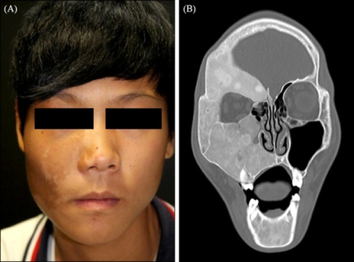
Author:Jung KE, Lee JH, Kim TY,Department of Dermatology, Eulji University Hospital, Daejeon, Korea (Openi/National Library of Medicine) Source:https://openi.nlm.nih.gov/detailedresult?img=PMC4198596_ad-26-639-g001&query=Polyostotic%20fibrous%20dysplasia&it=xg&req=4&npos=25 Description:F1: Solitary, brown patch with irregular border, throughout the patient's right cheek and upper eyelid with facial asymmetry. (B) Coronal computed tomography scan demonstrates heterogenous ground glass appearance at the right anterior skull, consistent with polyostotic fibrous dysplasia of the skull, right maxilla and mandible and narrowing of superior and inferior orbital canal, and compressing the right optic nerve.
File history
Click on a date/time to view the file as it appeared at that time.
| Date/Time | Thumbnail | Dimensions | User | Comment | |
|---|---|---|---|---|---|
| current | 20:45, 7 August 2021 | | 512 × 379 (288 KB) | Ozzie10aaaa (talk | contribs) | Author:Jung KE, Lee JH, Kim TY,Department of Dermatology, Eulji University Hospital, Daejeon, Korea (Openi/National Library of Medicine) Source:https://openi.nlm.nih.gov/detailedresult?img=PMC4198596_ad-26-639-g001&query=Polyostotic%20fibrous%20dysplasia&it=xg&req=4&npos=25 Description:F1: Solitary, brown patch with irregular border, throughout the patient's right cheek and upper eyelid with facial asymmetry. (B) Coronal computed tomography scan demonstrates heterogenous ground glass appearan... |
You cannot overwrite this file.
File usage
There are no pages that use this file.