File:PMC4210738 kjp-52-537-g002.png
PMC4210738_kjp-52-537-g002.png (512 × 226 pixels, file size: 157 KB, MIME type: image/png)
License
Attribution-NonCommercial 3.0 Unported (CC BY-NC 3.0)
Summary
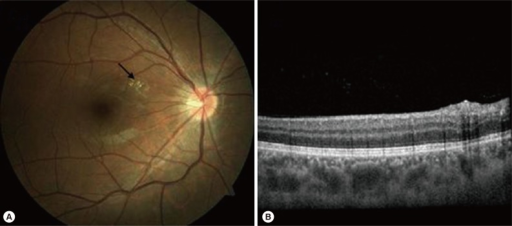
Author:Seong S, Moon D, Lee DK, Kim HE, Oh HS, Kim SH, Kwon OW, You YS,Nune Eye Hospital, Noon Building(Openi/National library of Medicine) Source:https://openi.nlm.nih.gov/detailedresult?img=PMC4210738_kjp-52-537-g002&query=Albendazole&it=xg&req=4&npos=5 Description:F2: Fundus photograph and optical coherence tomography of the right eye at 1 month after oral albendazole and triamcinolon therapy from day 13 of albendazole treatment. (A) Fundus photograph shows disappearance of the whitish epiretinal scar (arrow). (B) Decreased elevation of retinal surface and slightly remained posterior acoustic shadowing of the scar in optical coherence tomography.
File history
Click on a date/time to view the file as it appeared at that time.
| Date/Time | Thumbnail | Dimensions | User | Comment | |
|---|---|---|---|---|---|
| current | 22:41, 14 December 2021 | | 512 × 226 (157 KB) | Ozzie10aaaa (talk | contribs) | Author:Seong S, Moon D, Lee DK, Kim HE, Oh HS, Kim SH, Kwon OW, You YS,Nune Eye Hospital, Noon Building(Openi/National library of Medicine) Source:https://openi.nlm.nih.gov/detailedresult?img=PMC4210738_kjp-52-537-g002&query=Albendazole&it=xg&req=4&npos=5 Description:F2: Fundus photograph and optical coherence tomography of the right eye at 1 month after oral albendazole and triamcinolon therapy from day 13 of albendazole treatment. (A) Fundus photograph shows disappearance of the whitish epi... |
You cannot overwrite this file.
File usage
There are no pages that use this file.