File:PMC4215400 gr1.png
Jump to navigation
Jump to search
No higher resolution available.
PMC4215400_gr1.png (512 × 157 pixels, file size: 91 KB, MIME type: image/png)
Summary
| Description |
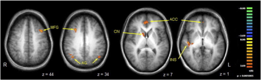
English: f0005: Contrast map of selected regions of BOLD activation that were significantly more active when vibrotactile stimuli were delivered to the symptomatic body part compared to the asymptomatic mirror region. Legend: anatomical images are derived from averages of T1-weighted scans from all participants and have been AC–PC aligned and transformed to Talairach space. MFG = middle frontal gyrus; AG = angular gyrus; CN = caudate nucleus; ACC = anterior cingulate cortex; INS = insula. See Table 1 for full list of activated regions. Per voxel cutoff of p < 0.005. |
| Date | |
| Source | https://openi.nlm.nih.gov/detailedresult?img=PMC4215400_gr1&query=Conversion%20disorder&it=xg&req=4&npos=3 |
| Author | Burke MJ, Ghaffar O, Staines WR, Downar J, Feinstein A |
Licensing
| This work is licensed under the Creative Commons Attribution 3.0 unported License.
Anyone may use this image for any purpose provided it is attributed as specified. |
 |
This file was uploaded with UploadWizard.
File history
Click on a date/time to view the file as it appeared at that time.
| Date/Time | Thumbnail | Dimensions | User | Comment | |
|---|---|---|---|---|---|
| current | 15:31, 30 May 2022 | 512 × 157 (91 KB) | Ozzie10aaaa (talk | contribs) | Uploaded a work by Burke MJ, Ghaffar O, Staines WR, Downar J, Feinstein A from https://openi.nlm.nih.gov/detailedresult?img=PMC4215400_gr1&query=Conversion%20disorder&it=xg&req=4&npos=3 with UploadWizard |
You cannot overwrite this file.
File usage
There are no pages that use this file.