File:PMC4228323 1471-2407-13-529-2.png
Jump to navigation
Jump to search

Size of this preview: 444 × 599 pixels. Other resolutions: 178 × 240 pixels | 512 × 691 pixels.
Original file (512 × 691 pixels, file size: 719 KB, MIME type: image/png)
Summary
| Description |
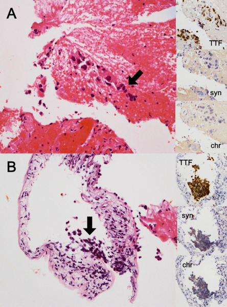
English: F2: Histopathology of combined small-cell lung cancer. A) Transbronchial lung biopsy from the primary lesion shows adenocarcinoma (hematoxylin and eosin staining, marked with arrow). The insets show that this tumor is positive for TTF-1 and negative for synaptophysin and chromogranin. B) Another slice from the same biopsy specimen shows small-cell lung cancer (hematoxylin and eosin staining, marked with arrow). The insets show that this tumor is positive for TTF-1, synaptophysin, and chromogranin. chr, chromogranin; syn, synaptophysin; TTF, TTF-1. |
| Date | |
| Source | https://openi.nlm.nih.gov/detailedresult?img=PMC4228323_1471-2407-13-529-2&query=Combined%20small-cell%20lung%20carcinoma&it=xg&req=4&npos=6 |
| Author | Takagi Y, Nakahara Y, Hosomi Y, Hishima T |
Licensing
{{subst:Custom license marker added by UW}} https://creativecommons.org/licenses/by/2.0/ Attribution 2.0 Generic (CC BY 2.0)
This file was uploaded with UploadWizard.
File history
Click on a date/time to view the file as it appeared at that time.
| Date/Time | Thumbnail | Dimensions | User | Comment | |
|---|---|---|---|---|---|
| current | 21:40, 1 September 2022 | | 512 × 691 (719 KB) | Ozzie10aaaa (talk | contribs) | Uploaded a work by Takagi Y, Nakahara Y, Hosomi Y, Hishima T from https://openi.nlm.nih.gov/detailedresult?img=PMC4228323_1471-2407-13-529-2&query=Combined%20small-cell%20lung%20carcinoma&it=xg&req=4&npos=6 with UploadWizard |
You cannot overwrite this file.
File usage
There are no pages that use this file.