File:PMC4231460 1752-1947-7-194-3.png
Jump to navigation
Jump to search
No higher resolution available.
PMC4231460_1752-1947-7-194-3.png (512 × 592 pixels, file size: 233 KB, MIME type: image/png)
Summary
| Description |
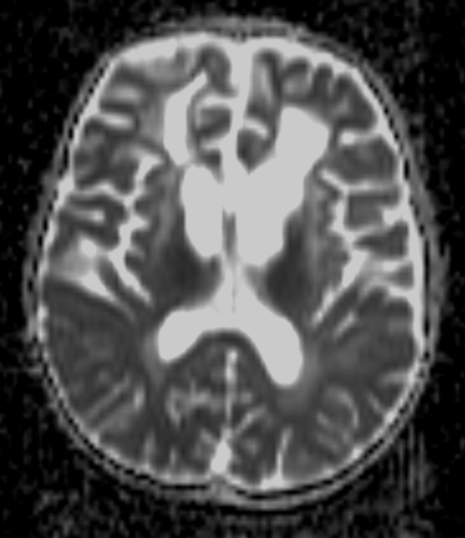
English: F3: Magnetic resonance imaging at 5 years, 4 months. a T2-weighted image shows increase in abnormal signals in the frontal lobe and volume loss in bilateral white matter and deep gray matter. b T2-weighted image demonstrates an increase in abnormal signals in the medulla oblongata. c Fluid attenuated inversion recovery image shows swelling and high intensity from the medulla oblongata to the top of the cervical spine. d, e Mild elevation in apparent diffusion coefficient values are recognized in affected areas. |
| Date | |
| Source | https://openi.nlm.nih.gov/detailedresult?img=PMC4231460_1752-1947-7-194-3&query=&req=4 |
| Author | Nishibayashi F, Kawashima M, Katada Y, Murakami N, Nozaki M |
Licensing
{{subst:Custom license marker added by UW}} https://creativecommons.org/licenses/by/2.0/ Attribution 2.0 Generic (CC BY 2.0)
This file was uploaded with UploadWizard.
File history
Click on a date/time to view the file as it appeared at that time.
| Date/Time | Thumbnail | Dimensions | User | Comment | |
|---|---|---|---|---|---|
| current | 19:03, 16 June 2022 | | 512 × 592 (233 KB) | Ozzie10aaaa (talk | contribs) | Uploaded a work by Nishibayashi F, Kawashima M, Katada Y, Murakami N, Nozaki M from https://openi.nlm.nih.gov/detailedresult?img=PMC4231460_1752-1947-7-194-3&query=&req=4 with UploadWizard |
You cannot overwrite this file.
File usage
There are no pages that use this file.