File:PMC4303217 mgg30002-0472-f5.png
PMC4303217_mgg30002-0472-f5.png (512 × 480 pixels, file size: 478 KB, MIME type: image/png)
License
Attribution 3.0 Unported (CC BY 3.0)
Summary
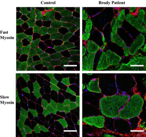
Author:Sambuughin N, Zvaritch E, Kraeva N, Sizova O, Sivak E, Dickson K, Weglinski M, Capacchione J, Muldoon S, Riazi S, Hamilton S, Brandom B, MacLennan DH ,Defense and Veterans Center for Integrated Pain Management, Henry M. Jackson Foundation Rockville, Department of Anesthesiology, Uniformed Services University Bethesda (Openi/National Library of Medicine) Source:https://openi.nlm.nih.gov/detailedresult?img=PMC4303217_mgg30002-0472-f5&query=&req=4 Description:fig05: Immunofluorescence staining for fast and slow myosins in the muscle of Brody patient (BP). Confocal microscopy images of transverse skeletal muscle sections stained with anti-fast (top) and anti-slow (bottom) myosin antibodies (green). WGA (red) stains connective tissue. DAPI (blue) counterstains nuclei. Increased fiber size variability and fiber hypertrophy is readily observed in both types of the BP muscle fibers. Bar, 100 μm.
File history
Click on a date/time to view the file as it appeared at that time.
| Date/Time | Thumbnail | Dimensions | User | Comment | |
|---|---|---|---|---|---|
| current | 16:06, 19 October 2021 | | 512 × 480 (478 KB) | Ozzie10aaaa (talk | contribs) | Author:Sambuughin N, Zvaritch E, Kraeva N, Sizova O, Sivak E, Dickson K, Weglinski M, Capacchione J, Muldoon S, Riazi S, Hamilton S, Brandom B, MacLennan DH ,Defense and Veterans Center for Integrated Pain Management, Henry M. Jackson Foundation Rockville, Department of Anesthesiology, Uniformed Services University Bethesda (Openi/National Library of Medicine) Source:https://openi.nlm.nih.gov/detailedresult?img=PMC4303217_mgg30002-0472-f5&query=&req=4 Description:fig05: Immunofluorescence sta... |
You cannot overwrite this file.
File usage
There are no pages that use this file.