File:PMC4320899 PRI2015-240505.002.png
PMC4320899_PRI2015-240505.002.png (512 × 114 pixels, file size: 135 KB, MIME type: image/png)
License
Attribution-NonCommercial 4.0 International (CC BY-NC 4.0)
Summary
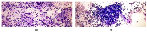
Author:Mehra P, Verma AK,Department Of Pathology, Employees State Insurance (ESI) Postgraduate Institute of Medical Sciences and Research and ESI Model Hospital(Openi/National Library of Medicine) Source:https://openi.nlm.nih.gov/detailedresult?img=PMC4320899_PRI2015-240505.002&query=Lymphomatous%20thyroiditis&it=xg&req=4&npos=10 Description:fig2: (a) Lymphocytic (Hashimoto) thyroiditis. Photomicrograph showing polymorphous lymphoid population (Smear, Giemsa, 400x magnification). (b) Lymphocytic (Hashimoto) thyroiditis. Photomicrograph showing lymphohistiocytic aggregates in lymphocytic (Hashimoto) thyroiditis (Smear, Giemsa, 400x magnification).
File history
Click on a date/time to view the file as it appeared at that time.
| Date/Time | Thumbnail | Dimensions | User | Comment | |
|---|---|---|---|---|---|
| current | 22:11, 21 December 2021 | 512 × 114 (135 KB) | Ozzie10aaaa (talk | contribs) | Author:Mehra P, Verma AK,Department Of Pathology, Employees State Insurance (ESI) Postgraduate Institute of Medical Sciences and Research and ESI Model Hospital(Openi/National Library of Medicine) Source:https://openi.nlm.nih.gov/detailedresult?img=PMC4320899_PRI2015-240505.002&query=Lymphomatous%20thyroiditis&it=xg&req=4&npos=10 Description:fig2: (a) Lymphocytic (Hashimoto) thyroiditis. Photomicrograph showing polymorphous lymphoid population (Smear, Giemsa, 400x magnification). (b) Lymphocy... |
You cannot overwrite this file.
File usage
There are no pages that use this file.