File:PMC4341013 10545 2014 9755 Fig3 HTML.png
Jump to navigation
Jump to search
No higher resolution available.
PMC4341013_10545_2014_9755_Fig3_HTML.png (512 × 276 pixels, file size: 53 KB, MIME type: image/png)
Summary
| Description |
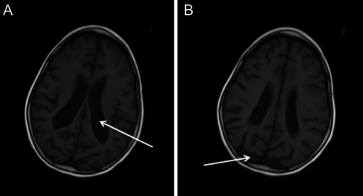
English: Fig3: a-b. Axial T2-weighted and T1-weighted cranial magnetic resonance of a patient with type I ADSL deficiency: arrows indicate symmetrical widening of the lateral ventricles, enlarged sulci of the brain cortex gyri, especially in the parieto-occipital lobes |
| Date | |
| Source | https://openi.nlm.nih.gov/detailedresult?img=PMC4341013_10545_2014_9755_Fig3_HTML&query=&req=4 |
| Author | Jurecka A, Zikanova M, Kmoch S, Tylki-Szymańska A |
Licensing
English: This file is licensed CC BY-NC 4.0
This file was uploaded with UploadWizard.
File history
Click on a date/time to view the file as it appeared at that time.
| Date/Time | Thumbnail | Dimensions | User | Comment | |
|---|---|---|---|---|---|
| current | 22:12, 24 June 2022 | | 512 × 276 (53 KB) | Ozzie10aaaa (talk | contribs) | Uploaded a work by Jurecka A, Zikanova M, Kmoch S, Tylki-Szymańska A from https://openi.nlm.nih.gov/detailedresult?img=PMC4341013_10545_2014_9755_Fig3_HTML&query=&req=4 with UploadWizard |
You cannot overwrite this file.
File usage
There are no pages that use this file.