File:PMC4389714 13256 2015 539 Fig3 HTML.png
Jump to navigation
Jump to search
No higher resolution available.
PMC4389714_13256_2015_539_Fig3_HTML.png (512 × 128 pixels, file size: 180 KB, MIME type: image/png)
Summary
| Description |
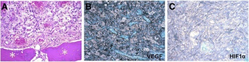
English: Fig3: Photomicrographs of retinal hemangioblastoma. (A) The retinal hemangioblastoma was composed of classical foamy (tumor) cells admixed with small capillaries. Osseous tissues (asterisks) are adjacent to the retinal hemangioblastoma. (B) The hemangioblastoma shows high expression of vascular endothelial growth factor (VEGF). (C) Hypoxia-inducible transcription factor 1α (HIF-1α) is expressed in the retinal hemangioblastoma. (A, hematoxylin and eosin, original magnification, ×200; B and C, avidin-biotin complex immunohistochemistry, original magnification, ×200). |
| Date | |
| Source | https://openi.nlm.nih.gov/detailedresult?img=PMC4389714_13256_2015_539_Fig3_HTML&query=Hemangioblastoma&it=xg&req=4&npos=3 |
| Author | Chen S, Chew EY, Chan CC |
Licensing
{{subst:Custom license marker added by UW}} https://creativecommons.org/publicdomain/zero/1.0/ CC0 1.0 Universal (CC0 1.0)Public Domain Dedication
&
Attribution 4.0 International (CC BY 4.0)
This file was uploaded with UploadWizard.
File history
Click on a date/time to view the file as it appeared at that time.
| Date/Time | Thumbnail | Dimensions | User | Comment | |
|---|---|---|---|---|---|
| current | 20:15, 12 October 2022 | 512 × 128 (180 KB) | Ozzie10aaaa (talk | contribs) | Uploaded a work by Chen S, Chew EY, Chan CC from https://openi.nlm.nih.gov/detailedresult?img=PMC4389714_13256_2015_539_Fig3_HTML&query=Hemangioblastoma&it=xg&req=4&npos=3 with UploadWizard |
You cannot overwrite this file.
File usage
There are no pages that use this file.