File:PMC4428802 pr-2013-01162k 0005.png
Jump to navigation
Jump to search
No higher resolution available.
PMC4428802_pr-2013-01162k_0005.png (512 × 569 pixels, file size: 480 KB, MIME type: image/png)
Summary
| Description |
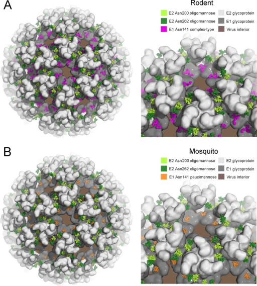
English: fig6: Models of the glycosylated alphavirus surface as derived from rodent(A) and mosquito (B) cells. The crystal structure of the CHKV E1–E2envelope glycoprotein complex was fitted into the Sindbis virus cryo-EMmap (PDB accession number 2XFB).12 E1 and E2 are shownin a surface representation in dark gray and light gray, respectively.Oligomannose-type glycans, presented by E2 at Asn200 and Asn262, areshown as light- and dark-green spheres, respectively (Man9GlcNAc2, from PDB accession code 2WAH(75)). At Asn141, glycan structures aremodeled as complex-type glycans for rodent-derived alphavirus (panelA; pink spheres, from PDB accession code 4BYH(74)) and paucimannose-typefor mosquito-derived alphavirus (panel B; orange spheres, from PDBaccession code 2WAH). |
| Date | |
| Source | https://openi.nlm.nih.gov/detailedresult?img=PMC4428802_pr-2013-01162k_0005&query=Alphavirus&it=xg&req=4&npos=6 |
| Author | Crispin M, Harvey DJ, Bitto D, Bonomelli C, Edgeworth M, Scrivens JH, Huiskonen JT, Bowden TA |
Licensing
English: This file is licensed CC BY-NC 4.0
This file was uploaded with UploadWizard.
File history
Click on a date/time to view the file as it appeared at that time.
| Date/Time | Thumbnail | Dimensions | User | Comment | |
|---|---|---|---|---|---|
| current | 22:42, 6 March 2023 | | 512 × 569 (480 KB) | Ozzie10aaaa (talk | contribs) | Uploaded a work by Crispin M, Harvey DJ, Bitto D, Bonomelli C, Edgeworth M, Scrivens JH, Huiskonen JT, Bowden TA from https://openi.nlm.nih.gov/detailedresult?img=PMC4428802_pr-2013-01162k_0005&query=Alphavirus&it=xg&req=4&npos=6 with UploadWizard |
You cannot overwrite this file.
File usage
There are no pages that use this file.