File:PMC4435298 crj-01-131-g001.png
PMC4435298_crj-01-131-g001.png (512 × 514 pixels, file size: 591 KB, MIME type: image/png)
License
Attribution-NonCommercial-NoDerivatives 4.0 International (CC BY-NC-ND 4.0)
Summary
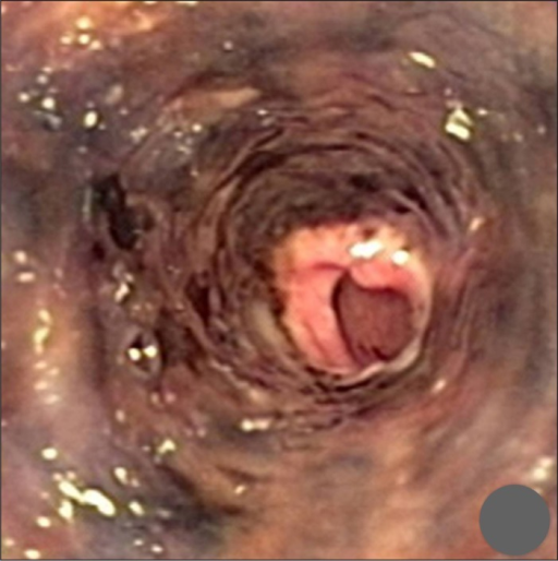
Author:Abed J, Mankal P, Judeh H, Kim S, Department of Medicine, Icahn School of Medicine, Mount Sinai St. Luke's and Roosevelt Hospitals(Openi/National Library of medicine) Source:https://openi.nlm.nih.gov/detailedresult?img=PMC4435298_crj-01-131-g001&query=&req=4 Description:F1: Endoscopy performed at the time of admission shows an ulcerated and necrotic esophageal mucosa starting 20 cm beyond the incisors to the gastroesophageal junction with normal gastric mucosa.
File history
Click on a date/time to view the file as it appeared at that time.
| Date/Time | Thumbnail | Dimensions | User | Comment | |
|---|---|---|---|---|---|
| current | 21:06, 8 December 2021 | | 512 × 514 (591 KB) | Ozzie10aaaa (talk | contribs) | Author:Abed J, Mankal P, Judeh H, Kim S, Department of Medicine, Icahn School of Medicine, Mount Sinai St. Luke's and Roosevelt Hospitals(Openi/National Library of medicine) Source:https://openi.nlm.nih.gov/detailedresult?img=PMC4435298_crj-01-131-g001&query=&req=4 Description:F1: Endoscopy performed at the time of admission shows an ulcerated and necrotic esophageal mucosa starting 20 cm beyond the incisors to the gastroesophageal junction with normal gastric mucosa. |
You cannot overwrite this file.
File usage
There are no pages that use this file.