File:PMC4461760 OMCL2015-725370.001.png
Jump to navigation
Jump to search
No higher resolution available.
PMC4461760_OMCL2015-725370.001.png (512 × 369 pixels, file size: 197 KB, MIME type: image/png)
Summary
| Description |
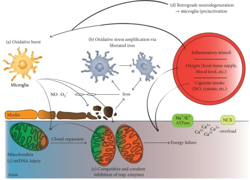
English: fig1: Oxidative stress-related mechanisms of tissue injury in multiple sclerosis. The temporal sequence and interconnection of cytotoxic events in MS may be different in individual lesions, stages of the disease, and different patients. Therefore, Figure 1 does not describe a timeline from events depicted in (a) to that in (d). (a) Microglia are activated by an unknown trigger pathology, the breakdown of the blood-brain barrier, and local and systemic inflammatory stimuli. Microglia activation itself may further impair the blood-brain barrier permeability. Microglia release nitric monoxide and superoxide molecules into the extracellular space. Nitric monoxide is uncharged and therefore penetrates lipid layers. Contrary to nitric monoxide, superoxide is unable to diffuse across biological membranes. It is rapidly converted into hydrogen peroxide, which, in contrast to superoxide, is able to diffuse into the extracellular environment. Additional amplification mechanisms involve microglia preactivation via axonal degeneration (d). (b) Iron is physiologically stored within the myelin sheets and liberated into the extracellular space upon demyelination. Extracellular iron amplifies oxidative stress as it travels between the ferrous and ferric states, inducing the production of highly reactive hydroxyl radicals. Iron is absorbed by microglia, which show histological signs of cell death under the high iron load and thus may release iron and initiate a second wave of oxidative stress. (c) Mitochondrial DNA (mtDNA) is vulnerable to free radical-mediated damage resulting in mtDNA deletions, which are found in neurons and axons of patients with multiple sclerosis. Mitochondria carrying such mutations are amplified by the clonal expansion in neurons. The mitochondrial respiratory chain is inhibited by covalent modifications caused by free radicals both competitively and irreversibly. A combination of these factors leads to energy failure via decreased ATP production. An important source of free radicals is the mitochondrial respiratory chain itself, particularly at low oxygen tension and reperfusion and in demyelinated axons. The exogenous factors, such as free radicals and cyanate delivered by smoking, inhibit mitochondrial function and cause demyelination in experimental conditions. Energy deficiency lowers Na+/K+-ATPase activity, resulting in the reverse operation of the Na+/Ca2+ exchanger (NCX) and thus increases Ca2+ levels. This event further activates the neurodegenerative and cell death pathways. |
| Date | |
| Source | https://openi.nlm.nih.gov/detailedresult?img=PMC4461760_OMCL2015-725370.001&query=multiple%20sclerosis&it=xg&req=4&npos=25 |
| Author | Haider L |
Licensing
English: This file is licensed CC BY-NC 4.0
This file was uploaded with UploadWizard.
File history
Click on a date/time to view the file as it appeared at that time.
| Date/Time | Thumbnail | Dimensions | User | Comment | |
|---|---|---|---|---|---|
| current | 16:44, 30 May 2022 | | 512 × 369 (197 KB) | Ozzie10aaaa (talk | contribs) | Uploaded a work by Haider L from https://openi.nlm.nih.gov/detailedresult?img=PMC4461760_OMCL2015-725370.001&query=multiple%20sclerosis&it=xg&req=4&npos=25 with UploadWizard |
You cannot overwrite this file.
File usage
There are no pages that use this file.