File:PMC4469251 13104 2015 1175 Fig1 HTML.png
Jump to navigation
Jump to search
Size of this preview: 480 × 600 pixels. Other resolutions: 192 × 240 pixels | 512 × 640 pixels.
Original file (512 × 640 pixels, file size: 227 KB, MIME type: image/png)
Summary
| Description |
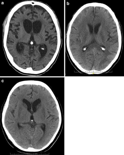
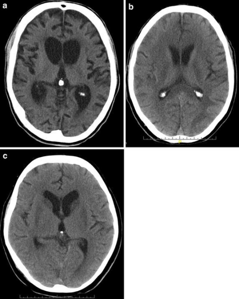
English: Typical CT scans of hypoxic brain damage patients. a Brain atrophy and hydrocephalus of a 44 y old male patient, 6 months after hypoxia. The patient was in a minimally conscious state (MCS). b Hypodense white matter changes of a 63 y old male patient, 2 weeks after hypoxic brain damage. The patient was in an unresponsive wakefulness syndrome (UWS). c Bilateral basal ganglia hypodensities of a 61 y old female patient, 6 weeks after hypoxic brain damage. The patient was in a MCS. |
| Date | |
| Source | https://openi.nlm.nih.gov/detailedresult?img=PMC4469251_13104_2015_1175_Fig1_HTML&query=brain%20damage&it=xg&req=4&npos=4 |
| Author | Heinz UE, Rollnik JD |
Licensing
{{subst:Custom license marker added by UW}} https://creativecommons.org/publicdomain/zero/1.0/ CC0 1.0 Universal (CC0 1.0) Public Domain Dedication
https://creativecommons.org/licenses/by/4.0/ Attribution 4.0 International (CC BY 4.0)
This file was uploaded with UploadWizard.
File history
Click on a date/time to view the file as it appeared at that time.
| Date/Time | Thumbnail | Dimensions | User | Comment | |
|---|---|---|---|---|---|
| current | 19:06, 14 May 2022 | | 512 × 640 (227 KB) | Ozzie10aaaa (talk | contribs) | Uploaded a work by Heinz UE, Rollnik JD from https://openi.nlm.nih.gov/detailedresult?img=PMC4469251_13104_2015_1175_Fig1_HTML&query=brain%20damage&it=xg&req=4&npos=4 with UploadWizard |
You cannot overwrite this file.
File usage
There are no pages that use this file.