File:PMC4496836 SNI-6-295-g003 (1).png
Jump to navigation
Jump to search
No higher resolution available.
PMC4496836_SNI-6-295-g003_(1).png (509 × 187 pixels, file size: 226 KB, MIME type: image/png)
Summary
| Description |
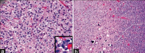
English: F3: Photomicrographs of anaplastic oligodendroglioma and arteriovenous malformation (AVM) histology (a) anaplastic oligodendroglioma showing uniform cellularity, uniformly round nuclei, and perinuclear halos. This section shows classic delicate microvasculature, although microvascular proliferation was seen in other areas. Inset highlights an atypical mitotic figure of which many were identified (H and E; original magnification, ×200). (b) Secondary features of oligodendroglioma include scattered microcalcifications (center of the image) and clonal nodules of heightened cellularity (right side of image) (H and E original magnification, ×100). (c) Irregular large vessels of the AVM with intervening cellular oligodendroglioma (*) (H and E original magnification, ×20). (d) Irregularly contoured vessels of the AVM filled with black granular embolization material. An intimal cushion is identified in one of the fibrotic vessel walls (*) (H and E original magnification, ×50) |
| Date | |
| Source | https://openi.nlm.nih.gov/detailedresult?img=PMC4496836_SNI-6-295-g003&query=Oligodendroglioma&it=xg&req=4&npos=1 |
| Author | Lai G, Muller KA, Carter BS, Chen CC |
Licensing
English: This file is licensed CC BY-NC-SA 3.0
This file was uploaded with UploadWizard.
File history
Click on a date/time to view the file as it appeared at that time.
| Date/Time | Thumbnail | Dimensions | User | Comment | |
|---|---|---|---|---|---|
| current | 18:44, 1 November 2022 | 509 × 187 (226 KB) | Ozzie10aaaa (talk | contribs) | Uploaded a work by Lai G, Muller KA, Carter BS, Chen CC from https://openi.nlm.nih.gov/detailedresult?img=PMC4496836_SNI-6-295-g003&query=Oligodendroglioma&it=xg&req=4&npos=1 with UploadWizard |
You cannot overwrite this file.
File usage
There are no pages that use this file.
.png&oldid=1248952)