File:PMC4518818 cln-70-08-544-g003.png
PMC4518818_cln-70-08-544-g003.png (512 × 344 pixels, file size: 355 KB, MIME type: image/png)
License
Attribution-NonCommercial 3.0 Unported (CC BY-NC 3.0)
Summary
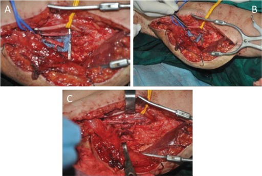
Author:Hou Y, Yang J, Yang Y, Qin B, Fu G, Li X, Gu L, Liu X, Zhu Q, Qi J , Sun Yat-sen University (Openi/National Library of medicine) Source:https://openi.nlm.nih.gov/detailedresult?img=PMC4518818_cln-70-08-544-g003&query=Brachial%20plexus%20injury&it=xg&req=4&npos=42 Description:f3-cln_70p544: Flow-through anastomosis of the T-shaped pedicle. A) The diameter of the profunda femoris segment is obviously larger than that of the nutrient artery of the gracilis. B) The brachial artery was resected, and the diameters of the segment profunda femoris and brachial artery were well matched. C) Interposed anastomosis to bridge the brachial artery. Two veins were anastomosed in direct end-to-end fashion.
File history
Click on a date/time to view the file as it appeared at that time.
| Date/Time | Thumbnail | Dimensions | User | Comment | |
|---|---|---|---|---|---|
| current | 17:28, 7 September 2021 | | 512 × 344 (355 KB) | Ozzie10aaaa (talk | contribs) | Author:Hou Y, Yang J, Yang Y, Qin B, Fu G, Li X, Gu L, Liu X, Zhu Q, Qi J , Sun Yat-sen University (Openi/National Library of medicine) Source:https://openi.nlm.nih.gov/detailedresult?img=PMC4518818_cln-70-08-544-g003&query=Brachial%20plexus%20injury&it=xg&req=4&npos=42 Description:f3-cln_70p544: Flow-through anastomosis of the T-shaped pedicle. A) The diameter of the profunda femoris segment is obviously larger than that of the nutrient artery of the gracilis. B) The brachial artery was rese... |
You cannot overwrite this file.
File usage
There are no pages that use this file.