File:PMC4575904 mv-v21-1093-f3.png
PMC4575904_mv-v21-1093-f3.png (512 × 167 pixels, file size: 180 KB, MIME type: image/png)
License
Attribution-NonCommercial-NoDerivs 3.0 Unported (CC BY-NC-ND 3.0)
Summary
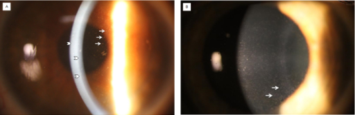
Author:Gee JA, Frausto RF, Chung DW, Tangmonkongvoragul C, Le DJ, Wang C, Han J, Aldave AJ,The Stein Eye Institute, David Geffen School of Medicine at UCLA (Openi/National Library of Medicine) Source:https://openi.nlm.nih.gov/detailedresult?img=PMC4575904_mv-v21-1093-f3&query=Fleck%20corneal%20dystrophy&it=xg&req=4&npos=2 Description:f3: Slit-lamp photomicrograph of two individuals with Fleck corneal dystrophy. A: In proband 1, punctate discrete opacities are visible in retroillumination (arrows), and a few larger opacities are seen in the posterior stroma in direct illumination (arrowheads). B: In proband 2, discrete punctate grayish-white opacities are seen diffusely distributed in the corneal stroma (arrows).
File history
Click on a date/time to view the file as it appeared at that time.
| Date/Time | Thumbnail | Dimensions | User | Comment | |
|---|---|---|---|---|---|
| current | 18:13, 12 August 2021 | 512 × 167 (180 KB) | Ozzie10aaaa (talk | contribs) | Author:Gee JA, Frausto RF, Chung DW, Tangmonkongvoragul C, Le DJ, Wang C, Han J, Aldave AJ,The Stein Eye Institute, David Geffen School of Medicine at UCLA (Openi/National Library of Medicine) Source:https://openi.nlm.nih.gov/detailedresult?img=PMC4575904_mv-v21-1093-f3&query=Fleck%20corneal%20dystrophy&it=xg&req=4&npos=2 Description:f3: Slit-lamp photomicrograph of two individuals with Fleck corneal dystrophy. A: In proband 1, punctate discrete opacities are visible in retroillumination (arr... |
You cannot overwrite this file.
File usage
There are no pages that use this file.