File:PMC4584485 13256 2015 696 Fig1 HTML.png
Jump to navigation
Jump to search
No higher resolution available.
PMC4584485_13256_2015_696_Fig1_HTML.png (473 × 328 pixels, file size: 159 KB, MIME type: image/png)
Summary
| Description |
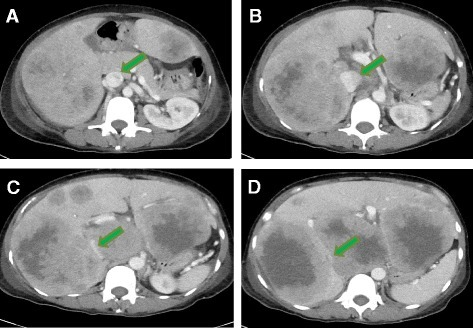
English: Fig1: Compression of the inferior vena cava by foci of hepatic metastases. Computed tomographic scan of the abdomen and pelvis with intravenous contrast shows that the inferior vena cava (green arrows) becomes progressively more compressed by the tumor superiorly to inferiorly. a Patent inferior vena cava at the level of the pancreas. d Near-complete compression of the inferior vena cava at a level of the superior portion of the kidneys. a-d Superiorly to inferiorly |
| Date | |
| Source | https://openi.nlm.nih.gov/detailedresult?img=PMC4584485_13256_2015_696_Fig1_HTML&query=&req=4 |
| Author | Patel SA |
Licensing
{{subst:Custom license marker added by UW}} https://creativecommons.org/licenses/by/4.0/ Attribution 4.0 International (CC BY 4.0)
&
CC0 1.0 Universal (CC0 1.0)Public Domain Dedication
This file was uploaded with UploadWizard.
File history
Click on a date/time to view the file as it appeared at that time.
| Date/Time | Thumbnail | Dimensions | User | Comment | |
|---|---|---|---|---|---|
| current | 23:27, 19 January 2023 | | 473 × 328 (159 KB) | Ozzie10aaaa (talk | contribs) | Uploaded a work by Patel SA from https://openi.nlm.nih.gov/detailedresult?img=PMC4584485_13256_2015_696_Fig1_HTML&query=&req=4 with UploadWizard |
You cannot overwrite this file.
File usage
There are no pages that use this file.