File:PMC4642028 kjim-30-6-938f1.png
Jump to navigation
Jump to search
No higher resolution available.
PMC4642028_kjim-30-6-938f1.png (512 × 141 pixels, file size: 98 KB, MIME type: image/png)
Summary
| Description |
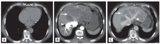
English: f1-kjim-30-6-938: Chest computed tomography (CT) reveals sparse pericardial calcification with eccentric wall thickening (A). Liver CT shows variable regions of low attenuation, often called the “nutmeg liver” presentation (B, C, arrows). Regurgitation of contrast material (B, arrowhead) to the intrahepatic veins, via the inferior vena cava, from the right atrium; with marked dilatation of the right, middle, and left hepatic veins (C, arrowhead) were noted. |
| Date | |
| Source | https://openi.nlm.nih.gov/detailedresult?img=PMC4642028_kjim-30-6-938f1&query=nutmeg%20liver&it=xg&req=4&npos=1 |
| Author | Shin KH, Joo HD, Song IH |
Licensing
English: This file is licensed CC BY-NC 3.0
This file was uploaded with UploadWizard.
File history
Click on a date/time to view the file as it appeared at that time.
| Date/Time | Thumbnail | Dimensions | User | Comment | |
|---|---|---|---|---|---|
| current | 18:21, 30 July 2022 | 512 × 141 (98 KB) | Ozzie10aaaa (talk | contribs) | Uploaded a work by Shin KH, Joo HD, Song IH from https://openi.nlm.nih.gov/detailedresult?img=PMC4642028_kjim-30-6-938f1&query=nutmeg%20liver&it=xg&req=4&npos=1 with UploadWizard |
You cannot overwrite this file.
File usage
There are no pages that use this file.