File:PMC4731993 13005 2016 102 Fig1 HTML.png
Jump to navigation
Jump to search
No higher resolution available.
PMC4731993_13005_2016_102_Fig1_HTML.png (472 × 340 pixels, file size: 370 KB, MIME type: image/png)
Summary
| Description |
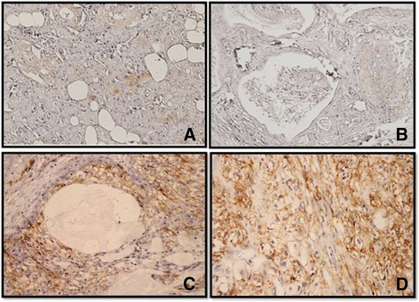
English: Fig1: Immunohistochemistry for CD44 in Mucoepidermoid carcinomas. a Normal salivary glands showed weak cytoplasmic stain in ductal epithelium and no immunoreactivity is seen in acinic cells. b No or mild CD44 expression was observed in the membrane of mucoepidermoid carcinoma cells (low grade type). c, d CD44 expression was strongly expressed in the membrane of the mucoepidermoid carcinoma cells (intermediate and high grade type) |
| Date | |
| Source | https://openi.nlm.nih.gov/detailedresult?img=PMC4731993_13005_2016_102_Fig1_HTML&query=Mucoepidermoid%20carcinoma&it=xg&req=4&npos=3 |
| Author | Binmadi N, Elsissi A, Elsissi N |
Licensing
{{subst:Custom license marker added by UW}} https://creativecommons.org/licenses/by/4.0/ Attribution 4.0 International (CC BY 4.0)
&
CC0 1.0 Universal (CC0 1.0)Public Domain Dedication
This file was uploaded with UploadWizard.
File history
Click on a date/time to view the file as it appeared at that time.
| Date/Time | Thumbnail | Dimensions | User | Comment | |
|---|---|---|---|---|---|
| current | 21:23, 8 January 2023 | | 472 × 340 (370 KB) | Ozzie10aaaa (talk | contribs) | Uploaded a work by Binmadi N, Elsissi A, Elsissi N from https://openi.nlm.nih.gov/detailedresult?img=PMC4731993_13005_2016_102_Fig1_HTML&query=Mucoepidermoid%20carcinoma&it=xg&req=4&npos=3 with UploadWizard |
You cannot overwrite this file.
File usage
There are no pages that use this file.