File:PMC4736587 CRIPA2016-9684910.004.png
PMC4736587_CRIPA2016-9684910.004.png (512 × 506 pixels, file size: 505 KB, MIME type: image/png)
License
Attribution-NonCommercial 4.0 International (CC BY-NC 4.0)
Summary
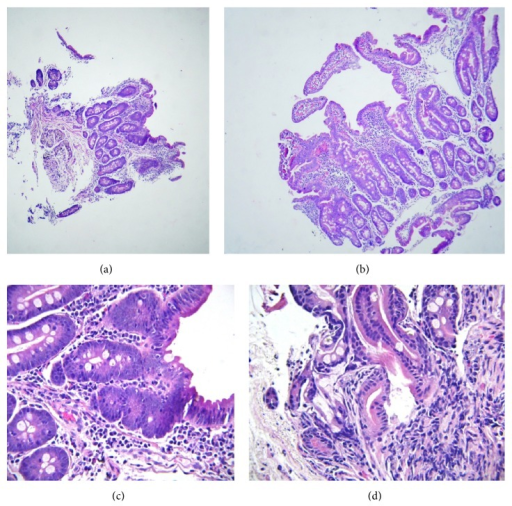
Author:Mahjoub FE, Imanzadeh F, Mahdavi Izadi S, Nahali Moghaddam A, Imam Khomeini Hospital Complex, Tehran University of Medical Sciences, Maternal, Fetal and Neonatal Research Center, Tehran University of Medical Sciences, Pediatric Nephrology Research Center, Tehran University of Medical Sciences, Roshan Azma Pathobiology Private Laboratory(Openi/National Library of Medicine)Source: https://openi.nlm.nih.gov/detailedresult?img=PMC4736587_CRIPA2016-9684910.004&query=&req=4 Description:fig4: Duodenal mucosa: (a) and (b) moderate to severe shortening of most villi (few rather tall villi are seen in picture (b)). (c) Focal superficial epithelial changes (poor gobletting, irregularity of lining cells) (intraepithelial lymphocytes: under 10/100). There is also mild to moderate infiltration of lymphoplasma cells and some eosinophils (3–7/100) in lamina propria. (d) Note crypt architectural changes.
File history
Click on a date/time to view the file as it appeared at that time.
| Date/Time | Thumbnail | Dimensions | User | Comment | |
|---|---|---|---|---|---|
| current | 22:53, 23 January 2022 | | 512 × 506 (505 KB) | Ozzie10aaaa (talk | contribs) | Author:Mahjoub FE, Imanzadeh F, Mahdavi Izadi S, Nahali Moghaddam A, Imam Khomeini Hospital Complex, Tehran University of Medical Sciences, Maternal, Fetal and Neonatal Research Center, Tehran University of Medical Sciences, Pediatric Nephrology Research Center, Tehran University of Medical Sciences, Roshan Azma Pathobiology Private Laboratory(Openi/National Library of Medicine)Source: https://openi.nlm.nih.gov/detailedresult?img=PMC4736587_CRIPA2016-9684910.004&query=&req=4 Description:fig4... |
You cannot overwrite this file.
File usage
There are no pages that use this file.