File:PMC4753938 medi-95-e2797-g001 (1) (1).png
Jump to navigation
Jump to search
No higher resolution available.
PMC4753938_medi-95-e2797-g001_(1)_(1).png (179 × 267 pixels, file size: 24 KB, MIME type: image/png)
Summary
| Description |
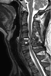
English: F1: Preoperative MRI. (A) Sagittal T2-weighted image of the cervical spine revealed large disc herniation at C4–C5 (arrow) and C5–C6 with severe spinal cord compression. The “Y-sign” (arrow head) was found. (B) Axial T2-weighted image of the cervical spine revealed large, central disc herniation at C4–C5 with severe spinal cord compression and surrounding edema. A “halo” of CSF isointensity surrounding the herniated disc was also demonstrated (arrow). CSF = cerebrospinal fluid, MRI = magnetic resonance imaging. |
| Date | |
| Source | https://openi.nlm.nih.gov/detailedresult?img=PMC4753938_medi-95-e2797-g001&query=Spinal%20cord%20compression&it=xg&req=4&npos=16 |
| Author | Yang HS, Oh YM, Eun JP |
Licensing
{{subst:Custom license marker added by UW}} https://creativecommons.org/licenses/by/4.0/ Attribution 4.0 International (CC BY 4.0)
This file was uploaded with UploadWizard.
File history
Click on a date/time to view the file as it appeared at that time.
| Date/Time | Thumbnail | Dimensions | User | Comment | |
|---|---|---|---|---|---|
| current | 15:46, 11 September 2022 | | 179 × 267 (24 KB) | Ozzie10aaaa (talk | contribs) | Uploaded a work by Yang HS, Oh YM, Eun JP from https://openi.nlm.nih.gov/detailedresult?img=PMC4753938_medi-95-e2797-g001&query=Spinal%20cord%20compression&it=xg&req=4&npos=16 with UploadWizard |
You cannot overwrite this file.
File usage
There are no pages that use this file.
_(1).png&oldid=1248535)