File:PMC4793739 12883 2016 555 Fig1 HTML (1).png
Jump to navigation
Jump to search
No higher resolution available.
PMC4793739_12883_2016_555_Fig1_HTML_(1).png (465 × 261 pixels, file size: 88 KB, MIME type: image/png)
Summary
| Description |
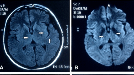
English: Fig1: Cranial MRI of our patient. Diffusion-weighted magnetic resonance imaging (DWI) (a) and the corresponding plane in fluid-attenuated inversion recovery (FLAIR) (b) showed bilateral hyper-intensity of the medial temporal lobe, insular lobe and basal ganglia (arrows). Repeated MRI were normal in February 28, 2015. (c, d) |
| Date | |
| Source | https://openi.nlm.nih.gov/detailedresult?img=PMC4793739_12883_2016_555_Fig1_HTML&query=Morvan%27s%20syndrome&it=xg&req=4&npos=2 |
| Author | Zhang L, Lu Q, Guan HZ, Mei JH, Ren HT, Liu MS, Peng B, Cui LY |
Licensing
{{subst:Custom license marker added by UW}} https://creativecommons.org/licenses/by/4.0/ Attribution 4.0 International (CC BY 4.0)
&
CC0 1.0 Universal (CC0 1.0)Public Domain Dedication
This file was uploaded with UploadWizard.
File history
Click on a date/time to view the file as it appeared at that time.
| Date/Time | Thumbnail | Dimensions | User | Comment | |
|---|---|---|---|---|---|
| current | 17:55, 11 September 2022 | | 465 × 261 (88 KB) | Ozzie10aaaa (talk | contribs) | Uploaded a work by Zhang L, Lu Q, Guan HZ, Mei JH, Ren HT, Liu MS, Peng B, Cui LY from https://openi.nlm.nih.gov/detailedresult?img=PMC4793739_12883_2016_555_Fig1_HTML&query=Morvan%27s%20syndrome&it=xg&req=4&npos=2 with UploadWizard |
You cannot overwrite this file.
File usage
There are no pages that use this file.
.png&oldid=1248538)