File:PMC4802527 gr2.png
PMC4802527_gr2.png (512 × 383 pixels, file size: 482 KB, MIME type: image/png)
License
Attribution-NonCommercial-NoDerivs 3.0 Unported (CC BY-NC-ND 3.0)
Summary
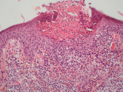
Author:Ropars N, Darrieux L, Tisseau L, Safa G ,Department of Dermatology, Centre Hospitalier de Saint-Brieuc(Openi/National Library of Medicine) Source:https://openi.nlm.nih.gov/detailedresult?img=PMC4802527_gr2&query=&req=4 Description:fig2: Acute generalized exanthematous pustulosis. A skin biopsy shows a subcorneal pustule filled with neutrophils. Note the presence of necrotic keratinocytes in the epidermis. Papillary dermal edema and superficial mixed infiltrate composed of lymphocytes and neutrophils can also be seen. (Hematoxylin-eosin stain; original magnification: ×200.)
File history
Click on a date/time to view the file as it appeared at that time.
| Date/Time | Thumbnail | Dimensions | User | Comment | |
|---|---|---|---|---|---|
| current | 19:33, 15 April 2022 | | 512 × 383 (482 KB) | Ozzie10aaaa (talk | contribs) | Author:Ropars N, Darrieux L, Tisseau L, Safa G ,Department of Dermatology, Centre Hospitalier de Saint-Brieuc(Openi/National Library of Medicine) Source:https://openi.nlm.nih.gov/detailedresult?img=PMC4802527_gr2&query=&req=4 Description:fig2: Acute generalized exanthematous pustulosis. A skin biopsy shows a subcorneal pustule filled with neutrophils. Note the presence of necrotic keratinocytes in the epidermis. Papillary dermal edema and superficial mixed infiltrate composed of lymphocytes... |
You cannot overwrite this file.
File usage
There are no pages that use this file.