File:PMC4820189 opth-10-527Fig1.png
Jump to navigation
Jump to search
No higher resolution available.
PMC4820189_opth-10-527Fig1.png (512 × 307 pixels, file size: 268 KB, MIME type: image/png)
Summary
| Description |
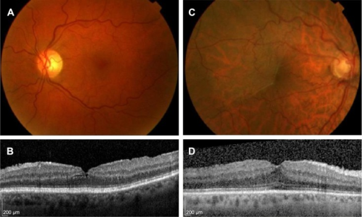
English: f1-opth-10-527: Epiretinal membrane examples.Notes: (A) Color fundus photograph demonstrating subtle cellophane macular reflex. (B) Spectralis OCT scan through the central fovea of (A) demonstrating a primary epiretinal membrane without significant retinal thickening (central foveal thickness of 274 μm) with an intact inner segment ellipsoid band. (C) Color fundus photograph demonstrating preretinal macular fibrosis. (D) Spectralis OCT scan through the central fovea of (C) demonstrating a primary epiretinal membrane with significant retinal thickening (central foveal thickness of 364 μm) with an intact inner segment ellipsoid band.Abbreviation: OCT, optical coherence tomography. |
| Date | |
| Source | https://openi.nlm.nih.gov/detailedresult?img=PMC4820189_opth-10-527Fig1&query=Epiretinal%20membrane&it=xg&req=4&npos=5 |
| Author | Stevenson W, Prospero Ponce CM, Agarwal DR, Gelman R, Christoforidis JB |
Licensing
English: This file is licensed CC BY-NC 3.0
&
This file was uploaded with UploadWizard.
File history
Click on a date/time to view the file as it appeared at that time.
| Date/Time | Thumbnail | Dimensions | User | Comment | |
|---|---|---|---|---|---|
| current | 18:55, 17 July 2022 | | 512 × 307 (268 KB) | Ozzie10aaaa (talk | contribs) | Uploaded a work by Stevenson W, Prospero Ponce CM, Agarwal DR, Gelman R, Christoforidis JB from https://openi.nlm.nih.gov/detailedresult?img=PMC4820189_opth-10-527Fig1&query=Epiretinal%20membrane&it=xg&req=4&npos=5 with UploadWizard |
You cannot overwrite this file.
File usage
There are no pages that use this file.