File:PMC4837931 40487 2013 1 Fig3 HTML.png
PMC4837931_40487_2013_1_Fig3_HTML.png (512 × 386 pixels, file size: 505 KB, MIME type: image/png)
License
Attribution-NonCommercial 4.0 International (CC BY-NC 4.0)
Summary
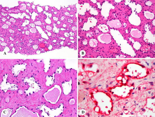
Author:Spencer KR, Miettinen MM, Maki RG, Mehnert JM,Department of Medicine, Rutgers University-Robert Wood Johnson Medical School (Openi/National Library of Medicine) Source:https://openi.nlm.nih.gov/detailedresult?img=PMC4837931_40487_2013_1_Fig3_HTML&query=Lymphangiomatosis&it=xg&req=4&npos=1 Description:Fig3: Lymphangiomatosis involving spleen and liver. a, b Lymphangiomatosis involving the spleen shows parenchymal replacement by vascular proliferation. The lumina are lined by attenuated endothelial cells and contain variably proteinaceous material. c Lymphangiomatosis involving the liver contains vascular profiles in fibrous stroma. Some profiles have vacuolated inward protuberant (“hobnail”) endothelia. d The endothelial cells are immunohistochemically positive for podoplanin (red chromogen)
File history
Click on a date/time to view the file as it appeared at that time.
| Date/Time | Thumbnail | Dimensions | User | Comment | |
|---|---|---|---|---|---|
| current | 20:01, 19 April 2022 | | 512 × 386 (505 KB) | Ozzie10aaaa (talk | contribs) | Author:Spencer KR, Miettinen MM, Maki RG, Mehnert JM,Department of Medicine, Rutgers University-Robert Wood Johnson Medical School (Openi/National Library of Medicine) Source:https://openi.nlm.nih.gov/detailedresult?img=PMC4837931_40487_2013_1_Fig3_HTML&query=Lymphangiomatosis&it=xg&req=4&npos=1 Description:Fig3: Lymphangiomatosis involving spleen and liver. a, b Lymphangiomatosis involving the spleen shows parenchymal replacement by vascular proliferation. The lumina are lined by attenuated... |
You cannot overwrite this file.
File usage
There are no pages that use this file.