File:PMC4851687 10545 2016 9929 Fig1 HTML.png
PMC4851687_10545_2016_9929_Fig1_HTML.png (512 × 410 pixels, file size: 199 KB, MIME type: image/png)
License
Attribution 4.0 International (CC BY 4.0)
Summary
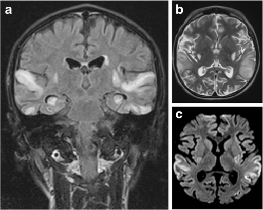
Author:Pittet MP, Idan RB, Kern I, Guinand N, Van HC, Toso S, Fluss J,Pediatric Neurology Unit, Pediatric Subspecialties Service, Children's Hospital, Geneva University Hospitals (Openi/National Library of Medicine) Source:https://openi.nlm.nih.gov/detailedresult?img=PMC4851687_10545_2016_9929_Fig1_HTML&query=MELAS%20syndrome&it=xg&req=4&npos=11 Description:Fig1: Brain MRI scan 72 h after onset of symptoms. a Coronal FLAIR image shows symmetrical high signal intensities lesions in multiple arterial territories. b Axial T2-weighted image demonstrating high signal cortical and subcortical lesions bilaterally in the edematous superior temporal gyri. c Axial diffusion-weighted image (DWI) demonstrating high signal areas in the same regions
File history
Click on a date/time to view the file as it appeared at that time.
| Date/Time | Thumbnail | Dimensions | User | Comment | |
|---|---|---|---|---|---|
| current | 20:13, 7 April 2022 | | 512 × 410 (199 KB) | Ozzie10aaaa (talk | contribs) | Author:Pittet MP, Idan RB, Kern I, Guinand N, Van HC, Toso S, Fluss J,Pediatric Neurology Unit, Pediatric Subspecialties Service, Children's Hospital, Geneva University Hospitals (Openi/National Library of Medicine) Source:https://openi.nlm.nih.gov/detailedresult?img=PMC4851687_10545_2016_9929_Fig1_HTML&query=MELAS%20syndrome&it=xg&req=4&npos=11 Description:Fig1: Brain MRI scan 72 h after onset of symptoms. a Coronal FLAIR image shows symmetrical high signal intensities lesions in multiple ar... |
You cannot overwrite this file.
File usage
There are no pages that use this file.