File:PMC4862290 JPN-11-56-g001.png
PMC4862290_JPN-11-56-g001.png (512 × 193 pixels, file size: 175 KB, MIME type: image/png)
License
Attribution-NonCommercial-ShareAlike 3.0 Unported (CC BY-NC-SA 3.0)
Summary
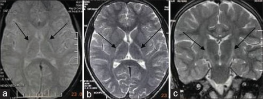
Author:Kumar S, Aga P, Gupta A, Kohli N , Department of Radiodiagnosis, King George's Medical University (Openi/National Library of Medicine) Source: https://openi.nlm.nih.gov/detailedresult?img=PMC4862290_JPN-11-56-g001&query=Juvenile%20primary%20lateral%20sclerosis&it=xg&req=4&npos=14 Description:F1: (a) T1-weighted imaging, black arrow shows hyperintensity in the posterior limb of internal capsule (b) T2-weighted imaging, black arrow shows hyperintensity in the posterior limb of bilateral internal capsule (c) T2-weighted imaging, black arrow shows hyperintensity in the posterior bilateral internal capsule and midbrain (corticospinal tract) with classical wine glass appearance
File history
Click on a date/time to view the file as it appeared at that time.
| Date/Time | Thumbnail | Dimensions | User | Comment | |
|---|---|---|---|---|---|
| current | 20:31, 12 August 2021 | 512 × 193 (175 KB) | Ozzie10aaaa (talk | contribs) | Author:Kumar S, Aga P, Gupta A, Kohli N , Department of Radiodiagnosis, King George's Medical University (Openi/National Library of Medicine) Source: https://openi.nlm.nih.gov/detailedresult?img=PMC4862290_JPN-11-56-g001&query=Juvenile%20primary%20lateral%20sclerosis&it=xg&req=4&npos=14 Description:F1: (a) T1-weighted imaging, black arrow shows hyperintensity in the posterior limb of internal capsule (b) T2-weighted imaging, black arrow shows hyperintensity in the posterior limb of bilateral... |
You cannot overwrite this file.
File usage
There are no pages that use this file.