File:PMC4885118 13256 2016 928 Fig1 HTML.png
Jump to navigation
Jump to search
No higher resolution available.
PMC4885118_13256_2016_928_Fig1_HTML.png (472 × 306 pixels, file size: 155 KB, MIME type: image/png)
Summary
| Description |
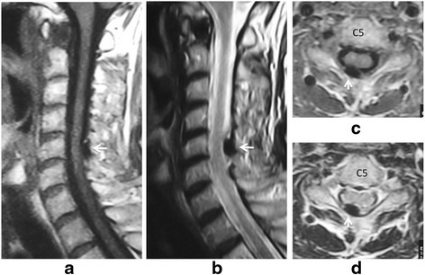
English: Fig1: Sagittal T1-weighted imaging (a) and T2-weighted imaging (b), and axial T1-weighted imaging (c) and T2WI (d) magnetic resonance imaging on initial admission. Low signal intensity (white arrow) on both T1-weighted imaging (a, c) and T2-weighted imaging (b, d) at interlaminar space of C5-C6 and C6-C7 is seen on the sagittal image (a, b) and axial image (c, d) |
| Date | |
| Source | https://openi.nlm.nih.gov/detailedresult?img=PMC4885118_13256_2016_928_Fig1_HTML&query=&req=4 |
| Author | Kobayashi T, Miyakoshi N, Abe T, Abe E, Kikuchi K, Noguchi H, Konno N, Shimada Y |
Licensing
{{subst:Custom license marker added by UW}} https://creativecommons.org/licenses/by/4.0/ Attribution 4.0 International (CC BY 4.0)
&
CC0 1.0 Universal (CC0 1.0)Public Domain Dedication
This file was uploaded with UploadWizard.
File history
Click on a date/time to view the file as it appeared at that time.
| Date/Time | Thumbnail | Dimensions | User | Comment | |
|---|---|---|---|---|---|
| current | 22:10, 10 March 2023 | | 472 × 306 (155 KB) | Ozzie10aaaa (talk | contribs) | Uploaded a work by Kobayashi T, Miyakoshi N, Abe T, Abe E, Kikuchi K, Noguchi H, Konno N, Shimada Y from https://openi.nlm.nih.gov/detailedresult?img=PMC4885118_13256_2016_928_Fig1_HTML&query=&req=4 with UploadWizard |
You cannot overwrite this file.
File usage
There are no pages that use this file.