File:PMC4890819 cabr-10-137-g003.png
PMC4890819_cabr-10-137-g003.png (512 × 255 pixels, file size: 112 KB, MIME type: image/png)
License
Attribution-NonCommercial-NoDerivatives 4.0 International (CC BY-NC-ND 4.0)
Summary
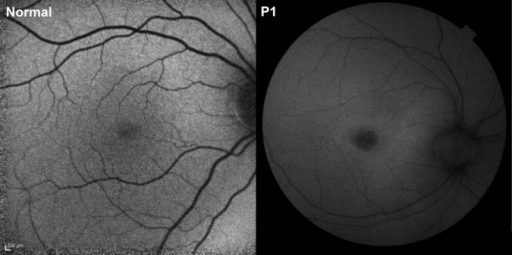
Author:Hansen MS, Hove MN, Jensen H, Larsen M ,Kennedy Center Eye Clinic, Glostrup Hospital, Department of Ophthalmology, Glostrup Hospital, and ‡Faculty of Health and Medical Sciences, University of Copenhagen (Openi/National Library of Medicine) Source:https://openi.nlm.nih.gov/detailedresult?img=PMC4890819_cabr-10-137-g003&query=Neuronal%20ceroid%20lipofuscinosis&it=xg&req=4&npos=4 Description:F3: Autofluorescence fundus photographs from a healthy 7-year-old girl (left) and from Patient 1 (right) with ceroid neuronal lipofuscinosis. The patient had a comparatively dark fovea and a normal or diffusely reduced autofluorescence in the rest of the fundus. Although the scale of the images is different, it is also apparent that the retinal vessels are thinner than normal in the patient.
File history
Click on a date/time to view the file as it appeared at that time.
| Date/Time | Thumbnail | Dimensions | User | Comment | |
|---|---|---|---|---|---|
| current | 15:07, 20 October 2021 | | 512 × 255 (112 KB) | Ozzie10aaaa (talk | contribs) | Author:Hansen MS, Hove MN, Jensen H, Larsen M ,Kennedy Center Eye Clinic, Glostrup Hospital, Department of Ophthalmology, Glostrup Hospital, and ‡Faculty of Health and Medical Sciences, University of Copenhagen (Openi/National Library of Medicine) Source:https://openi.nlm.nih.gov/detailedresult?img=PMC4890819_cabr-10-137-g003&query=Neuronal%20ceroid%20lipofuscinosis&it=xg&req=4&npos=4 Description:F3: Autofluorescence fundus photographs from a healthy 7-year-old girl (left) and from Patient 1... |
You cannot overwrite this file.
File usage
There are no pages that use this file.