File:PMC4899564 gr1.png
PMC4899564_gr1.png (512 × 122 pixels, file size: 52 KB, MIME type: image/png)
License
Attribution-NonCommercial-NoDerivatives 4.0 International (CC BY-NC-ND 4.0)
Summary
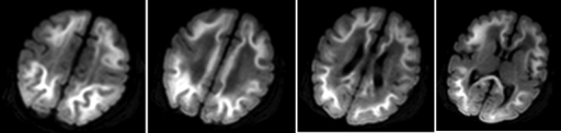
Author:Chang W, Gupta N, Duane D, Barnes P, Yeom K,Gundersen Lutheran Medical Foundation,Stanford University (Openi/National Library of Medicine) Source:https://openi.nlm.nih.gov/detailedresult?img=PMC4899564_gr1&query=Methylmalonic%20acidemia&it=xg&req=4&npos=2 Description:fig1: 6-day-old infant with methylmalonic acidemia. Sequential mean diffusion images derived from diffusion tensor imaging showed symmetric diffusion restriction in the subcortical white matter of bilateral cerebral hemispheres, with sparing of the peri-Rolandic regions. Note that there was no abnormal signal along the basal ganglia, and specifically along the globus pallidi.
File history
Click on a date/time to view the file as it appeared at that time.
| Date/Time | Thumbnail | Dimensions | User | Comment | |
|---|---|---|---|---|---|
| current | 22:49, 23 December 2021 | 512 × 122 (52 KB) | Ozzie10aaaa (talk | contribs) | Author:Chang W, Gupta N, Duane D, Barnes P, Yeom K,Gundersen Lutheran Medical Foundation,Stanford University (Openi/National Library of Medicine) Source:https://openi.nlm.nih.gov/detailedresult?img=PMC4899564_gr1&query=Methylmalonic%20acidemia&it=xg&req=4&npos=2 Description:fig1: 6-day-old infant with methylmalonic acidemia. Sequential mean diffusion images derived from diffusion tensor imaging showed symmetric diffusion restriction in the subcortical white matter of bilateral cerebral hemisp... |
You cannot overwrite this file.
File usage
There are no pages that use this file.