File:PMC4899878 gr4.png

Original file (512 × 681 pixels, file size: 917 KB, MIME type: image/png)
License
Attribution-NonCommercial-NoDerivatives 4.0 International (CC BY-NC-ND 4.0)
Summary
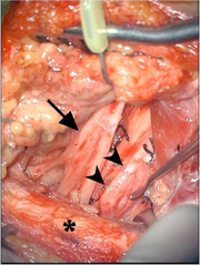
Author:Efrat Saraf Lavi,Allan D Levi,Erica K. Schallert, Andrew D. Brown, Michael D. Norenberg, Applebaum Diagnostic Imaging Center; the University of Miami Miller School of Medicine, Departments of Neurological Surgery, Orthopedics, and Rehabilitation the University of Miami Miller School of Medicine, Jackson Health System; the University of Miami Miller School of Medicine, Department of Pathology, Biochemistry, Neurology, and Neurosurgery, the University of Miami Miller School of Medicine(Openi/National Library of Medicine) Source:https://openi.nlm.nih.gov/detailedresult?img=PMC4899878_gr4&query=Intraneural%20perineurioma&it=xg&req=4&npos=4 Description:fig4: 18-year-old female with intraneural perineurioma. Intraoperative image shows an enlarged and fibrotic lower trunk of the brachial plexus (arrowheads) and the normal divisions of the upper trunk (arrow) located deep to the clavicle (asterisk) with the subclavian artery retracted inferomedially.
File history
Click on a date/time to view the file as it appeared at that time.
| Date/Time | Thumbnail | Dimensions | User | Comment | |
|---|---|---|---|---|---|
| current | 22:28, 23 January 2022 | | 512 × 681 (917 KB) | Ozzie10aaaa (talk | contribs) | Author:Efrat Saraf Lavi,Allan D Levi,Erica K. Schallert, Andrew D. Brown, Michael D. Norenberg, Applebaum Diagnostic Imaging Center; the University of Miami Miller School of Medicine, Departments of Neurological Surgery, Orthopedics, and Rehabilitation the University of Miami Miller School of Medicine, Jackson Health System; the University of Miami Miller School of Medicine, Department of Pathology, Biochemistry, Neurology, and Neurosurgery, the University of Miami Miller School of Medicine(... |
You cannot overwrite this file.
File usage
There are no pages that use this file.