File:PMC4928259 12884 2016 933 Fig6 HTML (1).png
Jump to navigation
Jump to search
No higher resolution available.
PMC4928259_12884_2016_933_Fig6_HTML_(1).png (198 × 198 pixels, file size: 27 KB, MIME type: image/png)
Summary
| Description |
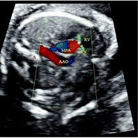
English: Fig6: Sonographic images of a 24-gestational-week fetus diagnosed with pulmonary valve atresia. Thickened pulmonary valves (indicated by arrows) were visualized in the right outflow tract view (a) via 2D. However, this anomaly was neglected by the sonographers under Protocol A because the valvular movement was difficult to visualize at times. However, color Doppler resolved this problem by visualizing the retrograde flow in the MPA (b), indicating pulmonary valve atresia. At the same time, no blood flow was detected entering the MPA across the thickened PV. AAO, ascending aorta; MPA, main pulmonary artery; PV, pulmonary valve; RV, right ventricle; S, spine |
| Date | |
| Source | https://openi.nlm.nih.gov/detailedresult?img=PMC4928259_12884_2016_933_Fig6_HTML&query=Pulmonary%20atresia&it=xg&req=4&npos=1 |
| Author | Zhang Y, Cai AL, Ren WD, Guo YJ, Zhang DY, Sun W, Wang Y, Wang L, Qin Y, Huang LP |
Licensing
{{subst:Custom license marker added by UW}} https://creativecommons.org/licenses/by/4.0/ Attribution 4.0 International (CC BY 4.0)
&
CC0 1.0 Universal (CC0 1.0)Public Domain Dedication
This file was uploaded with UploadWizard.
File history
Click on a date/time to view the file as it appeared at that time.
| Date/Time | Thumbnail | Dimensions | User | Comment | |
|---|---|---|---|---|---|
| current | 23:17, 10 October 2022 | | 198 × 198 (27 KB) | Ozzie10aaaa (talk | contribs) | Uploaded a work by Zhang Y, Cai AL, Ren WD, Guo YJ, Zhang DY, Sun W, Wang Y, Wang L, Qin Y, Huang LP from https://openi.nlm.nih.gov/detailedresult?img=PMC4928259_12884_2016_933_Fig6_HTML&query=Pulmonary%20atresia&it=xg&req=4&npos=1 with UploadWizard |
You cannot overwrite this file.
File usage
There are no pages that use this file.
.png&oldid=1248782)