File:PMC4965511 cpe-25-107-g001.png
PMC4965511_cpe-25-107-g001.png (512 × 205 pixels, file size: 69 KB, MIME type: image/png)
License
Attribution-NonCommercial-NoDerivs 3.0 Unported (CC BY-NC-ND 3.0)
Summary
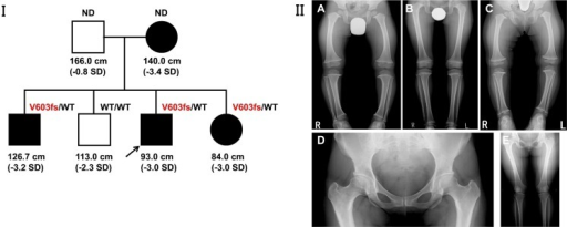
Author:Higuchi S, Takagi M, Shimomura S, Nishimura G, Hasegawa Y ,Department of Endocrinology and Metabolism, Tokyo Metropolitan Children's Medical Center (Openi/National Library of Medicine) Source:https://openi.nlm.nih.gov/detailedresult?img=PMC4965511_cpe-25-107-g001&query=Metaphyseal%20dysplasia&it=xg&req=4&npos=1 Description:fig_001: Characterization of the patient and his family. I: Pedigree ofthe family. II: Radiographs of the patient and his family. Radiographs of thepropositus at 3 yr of age (A), of the eldest brother at 11 yr of age (B), and of theyounger sister at 2 yr of age (C). The three siblings showed metaphysealirregularities in the hip and knee, coxa vara, and coxa magna. Both the propositus andhis younger siblings had bowlegs. Metaphyseal dysplasia was the most conspicuous inthe youngest sibling. Radiograph of the mother at 30 yr of age (D, E). The mothershowed coxa vara and short femoral necks. The long bones were somewhatstubby.
File history
Click on a date/time to view the file as it appeared at that time.
| Date/Time | Thumbnail | Dimensions | User | Comment | |
|---|---|---|---|---|---|
| current | 19:51, 7 August 2021 | 512 × 205 (69 KB) | Ozzie10aaaa (talk | contribs) | Author:Higuchi S, Takagi M, Shimomura S, Nishimura G, Hasegawa Y ,Department of Endocrinology and Metabolism, Tokyo Metropolitan Children's Medical Center (Openi/National Library of Medicine) Source:https://openi.nlm.nih.gov/detailedresult?img=PMC4965511_cpe-25-107-g001&query=Metaphyseal%20dysplasia&it=xg&req=4&npos=1 Description:fig_001: Characterization of the patient and his family. I: Pedigree ofthe family. II: Radiographs of the patient and his family. Radiographs of thepropositus at 3 y... |
You cannot overwrite this file.
File usage
There are no pages that use this file.