File:PMC5330223 jmmcr-03-5031-f002.png
PMC5330223_jmmcr-03-5031-f002.png (512 × 278 pixels, file size: 129 KB, MIME type: image/png)
License
Attribution 4.0 International (CC BY 4.0)
Summary
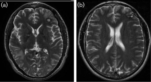
Author:Michael Eric Vollmer and Carol Glaser,Infectious Disease Doctors Medical Group,The Permanente Medical Group, Oakland Medical Center (Openi/National Library of Medicine) Source:https://openi.nlm.nih.gov/detailedresult?img=PMC5330223_jmmcr-03-5031-f002&query=&req=4 Description:F2: (a) HD#57 Axial T2 weighted magnetic resonance image (MRI) of brain demonstrating area of biopsy in left frontal region and oedema from mass in right temporal. Disease also noted with new satellite lesions seen in left frontal lobe and left parietal lobe. (b) MRI from two years into treatment with resolution of CNS lesions.
File history
Click on a date/time to view the file as it appeared at that time.
| Date/Time | Thumbnail | Dimensions | User | Comment | |
|---|---|---|---|---|---|
| current | 22:45, 6 November 2021 | | 512 × 278 (129 KB) | Ozzie10aaaa (talk | contribs) | Author:Michael Eric Vollmer and Carol Glaser,Infectious Disease Doctors Medical Group,The Permanente Medical Group, Oakland Medical Center (Openi/National Library of Medicine) Source:https://openi.nlm.nih.gov/detailedresult?img=PMC5330223_jmmcr-03-5031-f002&query=&req=4 Description:F2: (a) HD#57 Axial T2 weighted magnetic resonance image (MRI) of brain demonstrating area of biopsy in left frontal region and oedema from mass in right temporal. Disease also noted with new satellite lesions see... |
You cannot overwrite this file.
File usage
There are no pages that use this file.