File:PMC5366296 jvs-18-111-g002.png
PMC5366296_jvs-18-111-g002.png (512 × 168 pixels, file size: 82 KB, MIME type: image/png)
License
Attribution-NonCommercial 4.0 International (CC BY-NC 4.0)
Summary
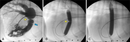
Author:Julia R. Treseder, SeungWoo Jung,Department of Clinical Sciences, College of Veterinary Medicine, Auburn University(Openi/National Library of Medicine) Source:https://openi.nlm.nih.gov/detailedresult?img=PMC5366296_jvs-18-111-g002&query=Pulmonic%20stenosis&it=xg&req=4&npos=3 Description:F2: Fluoroscopic images obtained before and during balloon dilation. (A) Right ventricular angiogram showing the pulmonary valve (blue arrow) and supravalvular pulmonic stenosis (yellow arrowhead). (B) Inflation of the balloon with diluted contrast solution across the stenotic lesion showing a discrete waist (yellow arrowhead) at the level of the supravalvular stenosis. (C) After full inflation of the balloon, complete loss of the waist is noted.
File history
Click on a date/time to view the file as it appeared at that time.
| Date/Time | Thumbnail | Dimensions | User | Comment | |
|---|---|---|---|---|---|
| current | 21:54, 2 January 2022 | 512 × 168 (82 KB) | Ozzie10aaaa (talk | contribs) | Author:Julia R. Treseder, SeungWoo Jung,Department of Clinical Sciences, College of Veterinary Medicine, Auburn University(Openi/National Library of Medicine) Source:https://openi.nlm.nih.gov/detailedresult?img=PMC5366296_jvs-18-111-g002&query=Pulmonic%20stenosis&it=xg&req=4&npos=3 Description:F2: Fluoroscopic images obtained before and during balloon dilation. (A) Right ventricular angiogram showing the pulmonary valve (blue arrow) and supravalvular pulmonic stenosis (yellow arrowhead). (B)... |
You cannot overwrite this file.
File usage
There are no pages that use this file.